Introduction to Spinal Cord Disorders and Imaging
Spinal cord disorders encompass a wide range of conditions, from traumatic injuries to degenerative diseases, that can significantly impact an individual’s mobility and quality of life. Diagnosing these disorders accurately is critical for effective treatment. Imaging techniques play a pivotal role in this diagnostic process, offering clear, detailed views of the spine, the spinal cord, and surrounding structures. Early detection and accurate assessment of spinal cord issues rely heavily on the appropriate use of advanced imaging tools. This helps healthcare professionals decide on the best course of treatment for the patient.
Medical disclaimer: This content is for general awareness and does not replace a doctor’s consultation. For diagnosis or treatment decisions, consult a qualified specialist.
Why Imaging is Crucial for Spinal Cord Diagnosis
Imaging is essential for diagnosing spinal cord disorders because it provides non-invasive, detailed insights into the spine's structure and function. Unlike physical examinations alone, imaging techniques can identify underlying issues such as herniated discs, spinal cord compression, or tumors. These imaging results help pinpoint the exact location, extent, and severity of a spinal issue, which is crucial for planning effective interventions. Without proper imaging, many spinal conditions could go undiagnosed or misdiagnosed, leading to inadequate treatment and potentially irreversible damage.

Understanding Spinal Cord Anatomy for Imaging
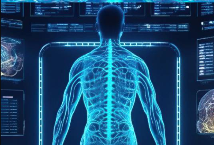
To interpret spinal cord imaging effectively, it is essential to have a thorough understanding of spinal anatomy. The spinal cord, housed within the vertebral column, extends from the brainstem to the lower back and is surrounded by protective structures such as vertebrae, ligaments, and cerebrospinal fluid. Imaging techniques allow clinicians to visualize not only the spinal cord but also the vertebral bones, discs, nerve roots, and blood vessels. This comprehensive view helps in diagnosing a wide range of spinal cord issues, including fractures, herniated discs, and spinal cord compression.
X-ray Imaging: The Basics and Its Role in Spinal Cord Diagnosis
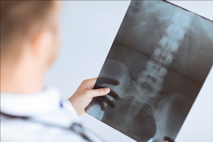
X-rays are one of the most commonly used imaging techniques to evaluate the spine. While they do not offer detailed images of soft tissues like the spinal cord, they are highly effective in assessing the bony structure of the spine. X-rays can reveal fractures, misalignments, or degenerative changes in the vertebrae, which may be indicative of spinal cord issues. For example, X-rays are often the first imaging modality used when trauma to the spine is suspected, helping healthcare professionals assess the bone integrity before further investigations are conducted using more advanced techniques.
MRI in Spinal Cord Imaging: A Comprehensive Overview
Magnetic Resonance Imaging (MRI) is one of the most powerful tools in diagnosing spinal cord disorders. MRI uses strong magnetic fields and radio waves to generate high-resolution images of the spinal cord and surrounding structures, including muscles, ligaments, and intervertebral discs. Unlike X-rays, MRI provides detailed soft tissue images, making it invaluable in identifying conditions such as herniated discs, tumors, infections, and multiple sclerosis. MRI can also be used to evaluate spinal cord injuries, allowing for precise assessment of the extent of damage.
CT Scans in Spinal Cord Injury and Pathology Detection
Computed Tomography (CT) scans combine X-ray images from multiple angles to produce detailed cross-sectional images of the spine. CT scans are particularly useful in detecting bony abnormalities, fractures, and other structural issues in the vertebral column. When combined with myelography, CT scans can also highlight issues involving the spinal cord and nerve roots. While MRI is often preferred for soft tissue evaluation, CT scans provide a quicker, more accessible alternative for detecting acute injuries and are sometimes used when MRI is unavailable.
The Role of Functional MRI in Spinal Cord Disorders
Functional MRI (fMRI) is an advanced technique that measures brain and spinal cord activity by detecting blood flow changes. In the context of spinal cord disorders, fMRI is particularly valuable in assessing how spinal cord injuries or pathologies affect motor and sensory functions. This technique is increasingly used to evaluate patients undergoing rehabilitation after spinal cord injuries, helping doctors gauge recovery and track functional improvements over time. fMRI can also be used in pre-surgical planning to avoid critical areas of the spinal cord during surgery.
PET and SPECT Imaging for Spinal Cord Evaluation
Positron Emission Tomography (PET) and Single-Photon Emission Computed Tomography (SPECT) are nuclear imaging techniques used to evaluate metabolic activity and blood flow within the spinal cord. These modalities are particularly useful in detecting tumors, infections, and other pathologies that affect spinal cord function. PET scans can provide insights into the physiological aspects of spinal cord disorders, while SPECT can help identify abnormalities in blood circulation, which is critical in diagnosing ischemia or vascular diseases affecting the spinal cord.
Ultrasound in Spinal Cord Injury Assessment: Pros and Cons
While ultrasound is most commonly used in the assessment of soft tissues and organs, its role in spinal cord imaging is limited. It can, however, be useful in evaluating superficial structures of the spine, especially in pediatric or trauma patients. Ultrasound may be used to guide procedures like spinal injections or to assess the flow of cerebrospinal fluid in cases of hydrocephalus. However, its ability to image the spinal cord itself is limited compared to other techniques like MRI and CT, and it is not typically used for deep or detailed spinal assessments.
Diffusion Tensor Imaging (DTI) in Spinal Cord Research
Diffusion Tensor Imaging (DTI) is an advanced form of MRI that provides insights into the microstructure of the spinal cord by mapping the movement of water molecules. DTI is particularly useful for assessing white matter integrity in the spinal cord, which can be damaged in conditions like multiple sclerosis, spinal cord injuries, and other neurodegenerative diseases. By analyzing the direction and speed of water diffusion, DTI helps clinicians understand how spinal cord damage affects communication between the brain and the rest of the body.
Magnetic Resonance Myelography: A Diagnostic Breakthrough
Magnetic Resonance Myelography (MRM) combines traditional MRI with the injection of contrast dye into the cerebrospinal fluid surrounding the spinal cord. This technique enhances the visibility of the spinal cord, nerve roots, and the surrounding tissues. MRM is particularly useful for detecting spinal cord compression, herniated discs, and other abnormalities that might not be as easily visible on a standard MRI. It is an excellent tool for diagnosing conditions like spinal stenosis or tumors that affect the spinal cord and nerve roots.
3D Imaging for Spinal Cord Pathologies
3D imaging provides highly detailed and accurate representations of the spinal cord and surrounding structures. By compiling multiple 2D imaging slices, 3D imaging allows for a comprehensive, three-dimensional view of the spine. This technology is especially useful in pre-surgical planning, as it helps surgeons identify the exact location and extent of spinal pathologies. It also aids in better understanding the spatial relationships between different structures in the spine, such as nerve roots, blood vessels, and the spinal cord itself.
CT Myelography: When It’s Necessary for Spinal Diagnosis
CT Myelography is a specialized imaging technique that combines CT scans with the injection of contrast dye into the spinal canal. This method helps visualize the spinal cord, nerve roots, and intervertebral discs, making it invaluable when MRI is contraindicated or unavailable. CT myelography is particularly effective for diagnosing spinal cord compression, tumors, and disc herniations that are not clearly visible on standard CT scans. It provides a detailed, clear image of the spinal cord and surrounding structures, which is crucial in planning surgical interventions.
The Role of Imaging in Spinal Cord Tumor Detection
Imaging plays a critical role in the detection and assessment of spinal cord tumors, which can be primary or metastatic. MRI is the gold standard for identifying tumors, as it provides clear, detailed images of soft tissues, allowing clinicians to distinguish between different types of tumors. In some cases, CT scans or myelography may be used to obtain additional details, especially when the tumor is located near bony structures. Early imaging detection of tumors is essential for developing an effective treatment plan, which may involve surgery, radiation therapy, or chemotherapy.
Spinal Stenosis Diagnosis Through Imaging Techniques
Spinal stenosis occurs when the spaces within the spine narrow, putting pressure on the spinal cord and nerves. Imaging techniques such as MRI are essential for diagnosing this condition, as they allow for the visualization of the spinal canal's narrowing and any associated issues like disc herniations or bone spurs. CT scans or myelography may also be used to get more detailed images in complex cases. Early diagnosis via imaging allows for timely intervention, which can help alleviate symptoms and prevent further nerve damage.

Spinal Cord Imaging for Degenerative Diseases: A Closer Look
Degenerative diseases such as osteoarthritis, disc degeneration, and spondylosis can lead to spinal cord compression and nerve impingement. MRI and CT scans are crucial for identifying the effects of these conditions, such as narrowing of the intervertebral discs or the formation of bone spurs. Imaging helps track the progression of these diseases and informs decisions regarding treatment options, such as conservative management, physical therapy, or surgery.
Imaging in Spinal Cord Congenital Abnormalities
Congenital abnormalities of the spinal cord, such as spina bifida or tethered cord syndrome, can often be identified early with the help of MRI. MRI provides high-resolution images of both the soft tissues and the vertebral column, making it the gold standard for evaluating congenital spinal issues. Early detection through imaging allows for timely intervention, which can prevent further complications and improve long-term outcomes for affected individuals.
The Importance of Early Diagnosis in Spinal Cord Disorders Using Imaging
Early diagnosis is critical for preventing permanent damage in many spinal cord disorders. Imaging techniques such as MRI and CT scans provide detailed insights that help healthcare providers identify issues at their onset, allowing for prompt treatment. For example, early detection of a herniated disc or spinal tumor can prevent further compression of the spinal cord, leading to better treatment outcomes and improved patient prognosis.
Imaging Techniques for Spinal Cord Inflammation and Demyelination
Conditions like multiple sclerosis (MS) and other inflammatory disorders that affect the spinal cord require specialized imaging. MRI is highly sensitive to changes in the spinal cord associated with demyelination, providing clear images of the affected areas. Advanced techniques, such as contrast-enhanced MRI, can detect active inflammation and track the development of lesions or plaques, aiding in diagnosis and treatment planning for these conditions.
Innovative Advances in Imaging for Spinal Cord Regeneration
Recent advancements in imaging technologies offer exciting potential for spinal cord regeneration research. Techniques like functional MRI and Diffusion Tensor Imaging (DTI) allow clinicians and researchers to track changes in the spinal cord structure and function following injury or disease. These innovations provide critical insights into how the spinal cord may regenerate or repair itself, which is key to developing new therapies aimed at restoring spinal cord function.
How Imaging Influences Treatment Plans for Spinal Cord Issues
Imaging influences treatment decisions by providing detailed information about the severity and location of spinal cord disorders. Depending on the findings, healthcare providers can choose the most appropriate treatment, whether it be conservative management, physical therapy, surgery, or a combination. Additionally, imaging is used to monitor the progress of treatment and assess the effectiveness of interventions, ensuring that patients receive the best care possible throughout their recovery journey.
Limitations of Current Spinal Cord Imaging Techniques
While imaging techniques like MRI and CT scans are invaluable for diagnosing spinal cord disorders, they do have limitations. For example, MRI can be expensive and may not be accessible in all healthcare settings. Additionally, certain conditions, such as small tumors or early-stage lesions, may not always be visible. These limitations often require the use of multiple imaging modalities in combination to ensure a complete and accurate assessment.
Comparing MRI and CT for Spinal Cord Diagnostics
MRI and CT scans each have distinct advantages when it comes to diagnosing spinal cord disorders. MRI is the preferred choice for evaluating soft tissue structures, such as the spinal cord, discs, and nerve roots, due to its high-resolution imaging. On the other hand, CT scans are excellent for visualizing bone abnormalities and offer faster imaging. Although MRI is preferred for most spinal cord evaluations, CT scans may be used in emergency situations or when MRI is contraindicated.
Innovative Surgical Tools Advancing Spinal Cord Procedures
Modern spinal cord surgeries are enhanced by tools like robotic-assisted systems, advanced imaging, and microsurgical instruments. These innovations allow for higher precision, minimal invasiveness, and faster recovery. They are crucial in procedures such as spinal decompression and fusion. Learn more about how advanced tools are improving spinal cord surgeries and patient outcomes.
Common Conditions Treated with Spinal Cord Surgery
Conditions like herniated discs, spinal stenosis, tumors, and injuries often require spinal cord surgery. These procedures aim to relieve pain, restore function, and prevent further damage. Surgical intervention is often life-changing for those with chronic pain or nerve-related issues. Explore the conditions effectively managed through surgery and the benefits they bring to patients.
The Future of AI and Machine Learning in Spinal Cord Imaging
Artificial Intelligence (AI) and machine learning are transforming the landscape of spinal cord imaging. These technologies can automate the analysis of imaging data, enhancing accuracy, reducing interpretation time, and identifying patterns that may go unnoticed by human radiologists. As AI evolves, it is expected to become an integral part of spinal cord diagnostics, potentially leading to earlier and more precise diagnoses.
The Role of Imaging in Spinal Cord Surgery Planning
Imaging is indispensable for preoperative planning in spinal cord surgeries. It helps surgeons pinpoint the exact location of abnormalities, such as herniated discs or spinal tumors, and plan the best surgical approach. 3D imaging provides additional precision, allowing for detailed visualization of the spine and its surrounding structures, which enhances surgical outcomes and reduces the risk of complications.
Best Spinal Cord Surgery in India
The Best Spinal Cord Surgery in India is performed by expert neurosurgeons who specialize in treating conditions affecting the spinal cord, using advanced techniques to ensure optimal patient outcomes and a personalized treatment plan based on individual health needs.
Best Spinal Cord Surgery Hospitals in India
The best spinal cord surgery hospitals in india are equipped with state-of-the-art technology and world-class facilities, providing comprehensive care from diagnosis to surgery and post-operative rehabilitation, ensuring a smooth recovery process for patients.
Spinal Cord Surgery Cost in India
When considering the spinal cord surgery cost in india, patients benefit from affordable, transparent pricing at leading hospitals, which offer cost-effective solutions without compromising the quality of care and surgical expertise.
Best Spinal Cord Surgery Doctors in India
The best spinal cord surgery doctors in india are highly experienced in treating complex spinal conditions, employing a patient-centric approach that ensures personalized care, precision in surgical techniques, and dedicated post-surgery follow-up to enhance recovery outcomes.
Minimally Invasive Spinal Cord Procedures Guided by Imaging
Advancements in minimally invasive spinal procedures are often guided by real-time imaging techniques such as fluoroscopy and intraoperative CT scans. These methods allow for precise navigation during spinal injections, biopsies, or surgeries, minimizing the need for large incisions. The use of imaging also reduces recovery times and lowers the risk of complications, making procedures safer and more efficient.
Patient-Centered Approaches in Spinal Cord Imaging
Patient-centered approaches in spinal cord imaging prioritize the comfort and safety of the patient throughout the diagnostic process. This involves selecting the most suitable imaging technique based on the patient's condition, medical history, and ability to tolerate the procedure. By considering the patient's needs, clinicians can ensure a more effective and less stressful imaging experience, ultimately leading to more accurate diagnoses and better overall care.
FAQs About the Role of Imaging Techniques in Diagnosing Spinal Cord Issues
What role does MRI play in diagnosing spinal cord disorders?
MRI is the gold standard for diagnosing spinal cord disorders. It provides high-resolution images of soft tissues, such as the spinal cord, nerve roots, and intervertebral discs, making it ideal for identifying issues like herniated discs, tumors, and spinal cord injuries.
How does CT scanning assist in spinal cord injury diagnosis?
CT scans are especially useful for detecting bone fractures or structural abnormalities that may impact the spinal cord. While MRI is better for soft tissue evaluation, CT scans can quickly identify injuries to the bony spine and help assess the extent of trauma.
What are the limitations of using ultrasound for spinal cord imaging?
Ultrasound is limited in its ability to assess the spinal cord directly. However, it is helpful for evaluating soft tissue structures around the spine and guiding procedures like injections. It is not typically used for comprehensive spinal cord diagnostics.
Can 3D imaging improve spinal cord diagnosis?
3D imaging enhances diagnostic accuracy by providing a detailed, three-dimensional view of the spine. It is especially helpful for planning surgeries and understanding the spatial relationships between structures within the spine, such as nerves and blood vessels.
What conditions can be detected through spinal cord MRI?
Spinal cord MRI can detect a wide variety of conditions, including herniated discs, spinal cord tumors, spinal stenosis, multiple sclerosis, and infections. It is highly effective in visualizing soft tissue abnormalities.
Is imaging necessary for spinal cord surgery?
Imaging is essential for planning spinal cord surgeries as it provides detailed maps of the affected areas. Surgeons rely on imaging to pinpoint the location and size of abnormalities and choose the optimal surgical approach.
What is the role of AI in spinal cord imaging?
AI helps enhance the accuracy and speed of spinal cord imaging by automating data analysis and identifying patterns that might be missed by human eyes. It has the potential to revolutionize diagnostic practices in the future.
How does CT myelography aid in spinal cord diagnostics?
CT myelography combines CT scans with contrast dye injected into the spinal canal to visualize the spinal cord, nerve roots, and other structures. It is particularly useful when MRI is not possible or does not provide sufficient detail.
What is Diffusion Tensor Imaging (DTI) and how is it used?
Diffusion Tensor Imaging (DTI) is an advanced MRI technique used to evaluate the microstructure of the spinal cord. It is particularly useful for detecting white matter changes and assessing damage in spinal cord injuries or diseases like multiple sclerosis.
Can imaging prevent spinal cord damage?
Imaging plays a vital role in early diagnosis and intervention, which can prevent spinal cord damage. Early detection of conditions like herniated discs or tumors allows for timely treatment, which can prevent further harm to the spinal cord.
Discover the Best Neurosurgery Hospitals and Neurosurgeons in India
When it comes to brain and spine care, choosing the right hospital and specialist is essential. We�ve highlighted the top neurosurgery hospitals and neurosurgeons across India to ensure you receive the best care available.
Top Neurosurgery Hospitals in India
Find the leading centers for brain and spine care:
Best Neurosurgeons in India
Meet the top specialists in brain and spine surgery:
Get more indepth information on Neurology treatments and their costs.
Conclusion
Your brain and spine health deserve the best care. Explore the links above to learn more about the top neurosurgery hospitals and neurosurgeons in India.
Discover the latest advances in minimally invasive spinal cord surgery. Learn how modern techniques, such as endoscopic procedures and robotic assistance, offer quicker recovery, reduced pain, and better outcomes for spinal cord conditions. Exploring Advances in Minimally Invasive Spinal Cord Surgery
Explore the anatomy of the spine and its influence on surgical procedures. Learn how the structure of vertebrae, discs, nerves, and muscles impacts the choice of spinal surgery techniques and outcomes for conditions like herniated discs and scoliosis. How Spine Anatomy Influences Surgical Approaches
Learn effective ways to minimize scarring after spinal cord surgery. Explore post-surgery care, including wound management, scar-reducing creams, massage techniques, and lifestyle changes that promote better healing and reduce visible scars. Tips to Minimize Scarring After Spinal Cord Surgery