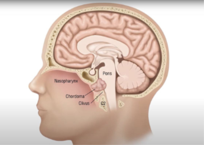
Introduction to Skull Base Conditions and Diagnosis
Skull base conditions refer to a diverse range of disorders that affect the complex area at the bottom of the skull, which houses critical structures such as the brainstem, cranial nerves, and major blood vessels. Diagnosing skull base conditions can be challenging due to the intricate anatomy and the variety of potential symptoms these conditions can present. Effective diagnosis often requires a combination of clinical evaluation, neurological examination, and advanced imaging techniques. Early and accurate diagnosis is essential for timely intervention, which can prevent complications and improve patient outcomes.
Overview of Common Skull Base Disorders
Common skull base disorders include both benign and malignant conditions. Tumors such as meningiomas, pituitary adenomas, and acoustic neuromas are among the most frequently encountered. These tumors can cause compression of surrounding structures, leading to symptoms such as headaches, vision problems, and hearing loss. Other skull base disorders include congenital abnormalities, like craniofacial deformities, vascular conditions like aneurysms, and traumatic injuries resulting from fractures or accidents.
Infections, such as skull base osteomyelitis, and inflammatory conditions, such as granulomatous diseases, can also affect this region. The complex nature of these conditions requires a multi-disciplinary approach to diagnosis and treatment, often involving neurosurgeons, otolaryngologists, and radiologists.

The Role of Clinical Evaluation in Diagnosing Skull Base Conditions
Clinical evaluation is a critical first step in diagnosing skull base conditions. The process typically begins with a detailed medical history and a thorough physical and neurological examination. Clinicians look for key symptoms such as persistent headaches, visual disturbances, balance issues, facial numbness, and difficulty hearing, which may indicate the involvement of structures at the skull base.
Neurological tests assessing cranial nerve function, coordination, and reflexes provide essential insights into which part of the skull base may be affected. Given the variety of conditions that can impact the skull base, clinical evaluation helps to narrow down potential diagnoses and guides the selection of appropriate imaging studies.
Neurological Symptoms: First Clues for Diagnosis
Neurological symptoms often provide the first indication of a skull base disorder. These symptoms can vary depending on the specific region of the skull base that is affected. For example, tumors or trauma near the anterior skull base may cause changes in smell (anosmia) or vision problems. Tumors affecting the middle or posterior skull base may lead to hearing loss, dizziness, or facial pain due to compression of cranial nerves.
Coordination problems, difficulty swallowing, and weakness in facial muscles may also indicate that the brainstem or cranial nerves are involved. Because these symptoms can overlap with other neurological conditions, recognizing them as early clues is crucial for prompting further diagnostic testing.
Imaging Techniques: The Key to Diagnosing Skull Base Conditions
Advanced imaging techniques are essential tools for diagnosing skull base conditions, providing detailed views of the complex anatomy. Magnetic resonance imaging (MRI) is often the preferred method, as it offers high-resolution images of soft tissues and can detect tumors, vascular abnormalities, and nerve involvement. MRI with contrast enhancement helps in identifying tumor borders and determining their relationship with surrounding structures.
Computed tomography (CT) scans are also frequently used, especially in cases of trauma, to assess fractures and bone abnormalities in the skull base. Angiography may be employed to visualize blood vessels when vascular conditions like aneurysms or arteriovenous malformations are suspected. In some cases, positron emission tomography (PET) scans or functional MRI may be used to further evaluate metabolic activity or brain function. Together, these imaging techniques provide the necessary information for a comprehensive diagnosis and aid in planning surgical or medical treatment.
MRI vs. CT Scans: When to Use Each for Skull Base Diagnosis
MRI (Magnetic Resonance Imaging) and CT (Computed Tomography) scans are essential diagnostic tools for assessing skull base conditions, but they are used differently based on the specific needs of each case. MRI provides superior soft tissue resolution, making it the preferred method for detecting tumors, nerve involvement, and abnormalities in brain structures. It is especially useful for diagnosing conditions like Chiari malformation or pituitary tumors.
CT scans excel in visualizing bony structures, making them ideal for assessing fractures, bone erosion, or trauma to the skull base. CT is often the first imaging choice for traumatic injuries and assessing the integrity of the skull base.
The Role of PET Scans in Skull Base Condition Diagnosis
PET scans (Positron Emission Tomography) are valuable for detecting metabolic activity in tissues, helping to identify malignant tumors at the skull base. PET scans are often used alongside MRI or CT to pinpoint cancerous growths and assess whether tumors have spread. They are particularly useful in evaluating cancers that have metastasized to the skull base or for differentiating between recurrent tumors and scar tissue after treatment.
Angiography for Diagnosing Vascular Conditions at the Skull Base
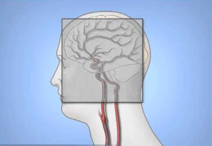
Angiography is crucial for diagnosing vascular conditions that affect the skull base, such as aneurysms, arteriovenous malformations, or tumors with a rich blood supply. Through the injection of contrast dye and the use of imaging techniques like MRI, CT, or conventional X-ray, angiography allows doctors to visualize the blood vessels and identify any abnormalities. This procedure is often used preoperatively to map out the vascular architecture around the skull base.

Endoscopy for Direct Visualization of Skull Base Structures
Endoscopy involves using a thin, flexible tube with a camera (endoscope) to directly visualize the structures at the skull base. This minimally invasive technique is often used for diagnosing and treating conditions such as pituitary tumors, CSF leaks, or sinus issues. Endoscopic skull base surgery allows for high-definition, magnified views of critical structures, enabling accurate diagnosis and targeted intervention.
Diagnosing Tumors: Biopsy Techniques and Procedures
When a skull base tumor is suspected, a biopsy may be required to confirm the diagnosis. Techniques can vary depending on the tumor's location. Minimally invasive approaches like needle biopsy can be used for accessible lesions, while more complex surgeries may be necessary for deep-seated tumors. The biopsy helps determine whether the tumor is benign or malignant, which informs the treatment plan.
Functional MRI (fMRI): Mapping Brain Activity for Skull Base Disorders
Functional MRI (fMRI) is used to map brain activity by measuring changes in blood flow. For skull base conditions affecting nearby brain structures, fMRI helps in planning surgery by identifying critical areas responsible for functions like speech, movement, or vision. This imaging technique is particularly useful in ensuring that vital brain functions are preserved during skull base surgery.
Electromyography (EMG) and Nerve Conduction Studies
Electromyography (EMG) and nerve conduction studies assess the electrical activity in muscles and nerves. These tests are useful in diagnosing nerve-related issues that may arise from skull base tumors, trauma, or cranial nerve compression. EMG measures muscle responses, while nerve conduction studies assess how well nerves transmit signals. Together, they provide critical information on the extent of nerve damage and guide treatment decisions.
The Importance of Early Diagnosis: Preventing Complications
Early diagnosis of skull base conditions is vital for preventing complications such as neurological deficits, vision or hearing loss, or life-threatening infections. Timely identification through imaging studies, genetic testing, and clinical evaluations allows for appropriate intervention before conditions progress, significantly improving patient outcomes and reducing the need for more invasive treatments later.
Genetic Testing: Identifying Congenital Skull Base Abnormalities
For congenital abnormalities of the skull base, genetic testing may be employed to identify underlying hereditary conditions. Genetic disorders like Ehlers-Danlos syndrome or other connective tissue diseases can predispose patients to skull base issues, such as Chiari malformation. Identifying these abnormalities early can guide treatment and inform family members of potential genetic risks.
Lumbar Puncture and Cerebrospinal Fluid (CSF) Analysis
Lumbar puncture, or spinal tap, is a procedure used to collect and analyze cerebrospinal fluid (CSF) to detect infections, CSF leaks, or increased intracranial pressure, which may be associated with skull base conditions. CSF analysis can help diagnose meningitis, certain types of tumors, or abnormalities in pressure that could indicate conditions like hydrocephalus or pseudotumor cerebri.
Diagnosing Infections at the Skull Base: Key Tests and Procedures
Diagnosing infections at the skull base requires a combination of imaging and laboratory tests. MRI with contrast is often used to detect soft tissue inflammation, abscesses, or infection spreading to surrounding structures. CT scans help identify bone involvement or erosion caused by the infection. Blood cultures, CSF analysis (via lumbar puncture), and other microbiological tests are essential for identifying the infectious agent. In some cases, a biopsy may be needed to confirm the type of infection, particularly for fungal or rare infections.
Neuro-Ophthalmic Evaluations: When Vision Is Affected
When skull base conditions affect the optic nerves or nearby structures, neuro-ophthalmic evaluations are critical. These assessments involve tests like visual field testing, visual acuity exams, and pupil reflex assessments to detect abnormalities in vision, optic nerve function, or eye movements. These evaluations help diagnose conditions such as pituitary tumors, meningiomas, or compressive lesions at the skull base affecting vision.
Vestibular Testing: Balance and Dizziness Diagnosis
For patients experiencing dizziness or balance issues due to skull base disorders, vestibular testing is crucial. Tests like electronystagmography (ENG), videonystagmography (VNG), and rotary chair testing assess inner ear and vestibular function, which may be affected by skull base tumors, trauma, or conditions like acoustic neuroma. These tests help identify the source of dizziness, allowing for targeted treatment.
Best Skull Base Surgery in India
The Best Skull Base Surgery in India is performed by expert neurosurgeons who utilize advanced techniques to ensure optimal outcomes for patients, offering a personalized treatment plan tailored to individual health needs.
Best Skull Base Surgery Hospitals in India
The best skull base surgery hospitals in india are equipped with cutting-edge technology and facilities, providing top-notch care, including pre-surgery consultations, surgical expertise, and post-operative recovery support to ensure a smooth patient journey.
Skull Base Surgery Cost in India
When considering the skull base surgery cost in india, patients benefit from affordable and transparent pricing at leading hospitals, which offer cost-effective treatment options without compromising the quality of care.
Best Skull Base Surgery Doctors in India
The best skull base surgery doctors in india are highly experienced in performing the procedure, utilizing a patient-centric approach that ensures personalized care, precise surgical techniques, and dedicated follow-up care to enhance recovery.
Multidisciplinary Approach: How Specialists Collaborate on Diagnosis
A multidisciplinary approach is essential for diagnosing skull base conditions due to the complexity and diversity of symptoms. Teams of specialists—including neurosurgeons, neurologists, ENT (ear, nose, and throat) surgeons, ophthalmologists, and radiologists—collaborate to interpret imaging studies, conduct neurological exams, and perform specialized tests. This collective effort ensures accurate diagnosis and comprehensive treatment planning.
Future Trends in Skull Base Condition Diagnostics
Advancements in diagnostic tools for skull base conditions are focused on improving precision and reducing invasiveness. Emerging technologies like 3D imaging, intraoperative MRI, and robot-assisted endoscopy are improving visualization and accuracy in diagnosis and surgery. Artificial intelligence (AI) is being integrated into imaging interpretation, allowing for more rapid detection of abnormalities. Additionally, genomic profiling is becoming more prominent, especially for identifying congenital skull base conditions and personalized treatment approaches.
FAQs About the Diagnostic Approaches for Skull Base Conditions
What are the most common diagnostic tools for skull base conditions?
Common tools include MRI, CT scans, PET scans, angiography, endoscopy, and biopsy.
How is MRI used to diagnose skull base disorders?
MRI provides high-resolution images of soft tissue, making it ideal for detecting tumors, infections, and nerve compression at the skull base.
When is a CT scan more appropriate than an MRI for diagnosing skull base conditions?
CT scans are preferred for evaluating bone structures, fractures, or bone erosion due to trauma, infections, or tumors.
What role does endoscopy play in diagnosing skull base conditions?
Endoscopy allows direct visualization of the skull base and is particularly useful for diagnosing and treating conditions like pituitary tumors or CSF leaks.
How are vascular conditions at the skull base diagnosed?
Vascular conditions are typically diagnosed using angiography, either through MRI, CT, or conventional methods, to visualize blood vessels and detect issues like aneurysms or arteriovenous malformations.
What tests are used to diagnose infections at the skull base?
Tests include MRI, CT scans, blood cultures, CSF analysis, and sometimes biopsy to identify the infectious agent and assess tissue or bone involvement.
How do doctors diagnose skull base tumors?
Skull base tumors are diagnosed using a combination of MRI, CT, PET scans, and sometimes biopsy to determine the type and extent of the tumor.
What is the role of nerve conduction studies in diagnosing skull base disorders?
Nerve conduction studies help assess the function of cranial nerves, particularly in cases where tumors, trauma, or compression at the skull base may cause nerve dysfunction.
How can genetic testing help in diagnosing skull base conditions?
Genetic testing can identify hereditary or congenital conditions that predispose individuals to skull base abnormalities, such as Ehlers-Danlos syndrome or Chiari malformation.
What are the latest advancements in diagnosing skull base disorders?
Advancements include 3D imaging, AI-enhanced imaging interpretation, minimally invasive biopsy techniques, and intraoperative MRI for more precise diagnosis and treatment.