Cardiac imaging plays a critical role in the diagnosis and management of Tetralogy of Fallot (TOF), a congenital heart defect characterized by four key abnormalities that affect normal blood flow. Early and accurate diagnosis is essential for planning the appropriate treatment, which typically involves surgery. Advances in cardiac imaging technologies have significantly improved the ability to detect and evaluate the severity of TOF, guiding clinicians in their decisions regarding surgical intervention and post-operative care.
Medical disclaimer: This content is for general awareness and does not replace a doctor’s consultation. For diagnosis or treatment decisions, consult a qualified specialist.
Understanding Tetralogy of Fallot and Its Diagnostic Challenges
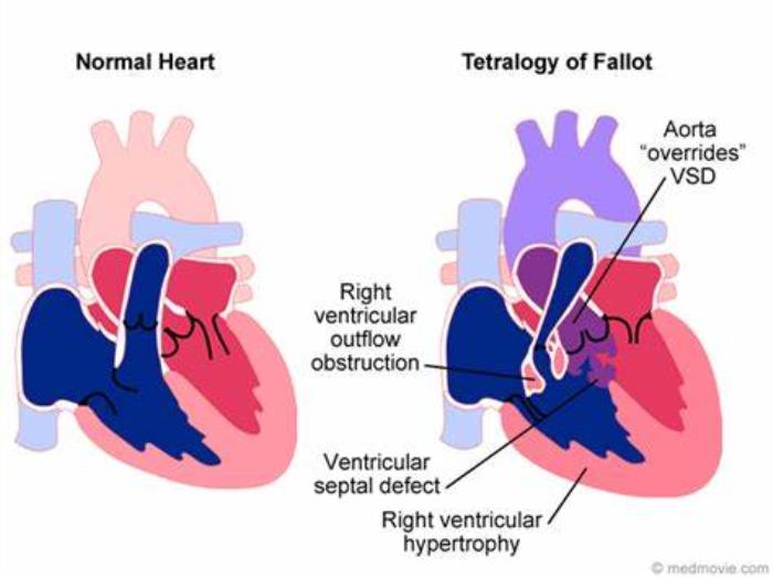
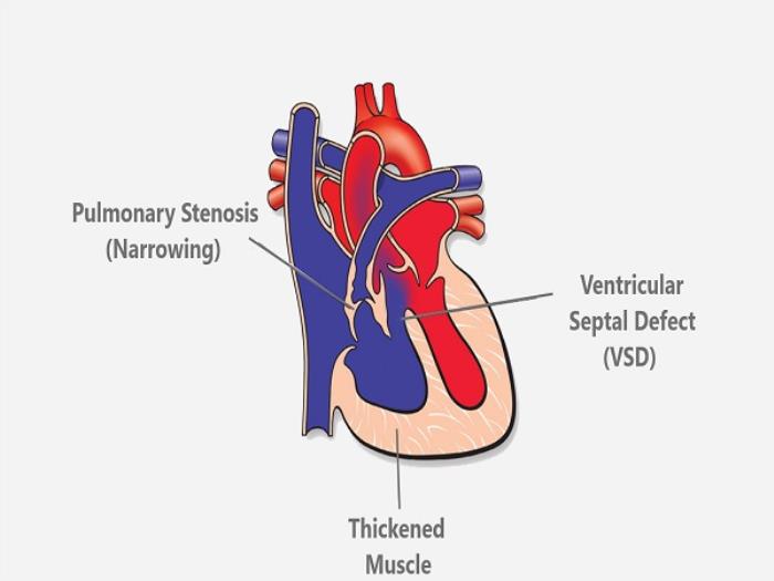
Tetralogy of Fallot involves a combination of structural heart defects, including a ventricular septal defect, pulmonary stenosis, an overriding aorta, and right ventricular hypertrophy. These abnormalities lead to reduced oxygen levels in the blood and cause cyanosis (a bluish tint to the skin), which is a hallmark of the condition. Diagnosing TOF can be challenging because the severity of symptoms varies, and some children may present with mild manifestations, making early detection crucial for optimal outcomes.
Traditional Imaging Methods for Tetralogy of Fallot Diagnosis
Historically, diagnosing Tetralogy of Fallot relied heavily on traditional imaging methods such as chest X-rays, electrocardiograms (ECGs), and standard echocardiography. While these techniques provided valuable information, they had limitations in terms of visualizing the intricate anatomical details of the heart, which are essential for assessing the extent of the defect and planning surgical intervention. Chest X-rays, for example, can indicate heart enlargement or pulmonary congestion, but they don’t provide detailed structural insights.

The Evolution of Cardiac Imaging Technologies for Tetralogy of Fallot
Over the years, advances in cardiac imaging technologies have revolutionized the ability to diagnose and manage Tetralogy of Fallot. The introduction of more sophisticated imaging modalities, such as 3D echocardiography, cardiac MRI, and CT scans, has provided clinicians with high-resolution images that allow for a more precise understanding of the heart’s anatomy and function. These tools offer detailed, dynamic views of the heart, improving both diagnosis and the planning of surgical procedures.
How Echocardiography Has Improved the Diagnosis of Tetralogy of Fallot
Echocardiography remains one of the most valuable and widely used imaging techniques in diagnosing Tetralogy of Fallot. Advances in echocardiographic technology, particularly with the advent of 3D echocardiography, have allowed for highly detailed imaging of the heart’s chambers, valves, and blood flow. This non-invasive, real-time imaging modality enables clinicians to assess the size and location of the ventricular septal defect, the degree of pulmonary stenosis, and the extent of right ventricular hypertrophy, all critical factors in managing TOF. The ability to visualize blood flow dynamics also helps determine the urgency of surgical intervention.
The Role of MRI in Accurate Diagnosis of Tetralogy of Fallot
Magnetic Resonance Imaging (MRI) has become an essential tool in diagnosing Tetralogy of Fallot, particularly in complex cases where echocardiography may provide limited information. MRI offers high-resolution images of the heart’s structure and function, enabling detailed visualization of the pulmonary arteries, right ventricle, and the aortic root. It is particularly useful for assessing the impact of pulmonary stenosis and determining the exact location and size of the ventricular septal defect. MRI is also valuable for preoperative planning, providing insights into the best surgical approach and post-operative prognosis.
Advancements in CT Imaging for Tetralogy of Fallot Diagnosis
Computed Tomography (CT) imaging has emerged as another valuable tool in diagnosing Tetralogy of Fallot, particularly for evaluating the aortic root, pulmonary arteries, and other vascular structures. Recent advancements in CT technology, such as improved image resolution and faster scan times, have made it possible to obtain detailed, three-dimensional images of the heart and surrounding vessels. CT imaging is particularly helpful in assessing vascular anomalies, surgical planning, and evaluating post-surgical outcomes. It provides a complementary tool to echocardiography and MRI, especially when detailed anatomical information is required to guide intervention.
Three-Dimensional Imaging: A Breakthrough in Tetralogy of Fallot Diagnosis
Three-dimensional (3D) imaging has revolutionized the diagnosis of Tetralogy of Fallot (TOF) by providing a detailed and accurate representation of the heart’s anatomy. This advanced imaging allows doctors to visualize the complex structural abnormalities associated with TOF, such as the ventricular septal defect, pulmonary stenosis, and overriding aorta, in a way that traditional imaging methods cannot. It enhances the accuracy of diagnosis, enables better planning for surgery, and aids in tracking the condition over time.
The Benefits of Cardiac Angiography in Visualizing Tetralogy of Fallot
Cardiac angiography plays a vital role in visualizing blood vessels and assessing the degree of pulmonary stenosis or obstruction in patients with Tetralogy of Fallot. This technique provides detailed images of the heart's blood flow and can help identify any associated congenital abnormalities, such as abnormal coronary artery connections, that may affect treatment plans. It is a crucial tool for planning interventions and monitoring the effectiveness of surgical repairs.
How Advanced Imaging Helps in Pre-Surgery Planning for Tetralogy of Fallot
Advanced imaging techniques such as 3D echocardiography and cardiac MRI allow for precise planning of surgeries for Tetralogy of Fallot. By offering detailed views of the heart's anatomy, including the size of defects, the position of the pulmonary artery, and the right ventricle's condition, imaging guides surgeons in determining the best approach for repair. These technologies help optimize surgical outcomes by providing an in-depth understanding of the structural challenges.
Role of Imaging in Assessing Pulmonary Valve and Right Ventricular Function
Imaging techniques, including MRI and echocardiography, are instrumental in assessing the function of the pulmonary valve and the right ventricle in patients with Tetralogy of Fallot. These tools can detect changes in pressure gradients, valve dysfunction, and ventricular enlargement, all of which are crucial in determining the necessity for further surgical interventions or interventions to manage pulmonary valve dysfunction and right ventricular pressures.
Integrating Imaging with Genetic Testing for a Comprehensive Diagnosis
When imaging is combined with genetic testing, a more comprehensive diagnosis of Tetralogy of Fallot can be made. Genetic testing can identify underlying genetic causes or syndromes that may contribute to the condition, while imaging provides real-time, structural details of the heart’s anatomy. Together, these tools help create a thorough understanding of the patient’s condition, which is essential for both diagnosis and tailoring a personalized treatment plan.

The Impact of Imaging Advances on Early Detection of Tetralogy of Fallot
Imaging advances, particularly high-resolution ultrasound and MRI, have significantly improved the early detection of Tetralogy of Fallot. With more precise imaging, abnormalities such as ventricular septal defects and pulmonary stenosis can be detected in fetal or early infant stages. Early detection enables timely interventions, such as surgical repair or catheter-based treatments, that can improve long-term outcomes for affected children.
How High-Resolution Imaging Improves Surgical Outcomes for Tetralogy of Fallot Patients
High-resolution imaging improves surgical outcomes for Tetralogy of Fallot patients by providing detailed, real-time information about the heart's structure and function. Surgeons can use advanced imaging to plan the surgery more accurately, avoid complications, and ensure that the repairs are done precisely. This level of detail enhances both the safety and effectiveness of the surgery, leading to better recovery and long-term health.
The Future of Cardiac Imaging: What’s Next for Tetralogy of Fallot Diagnosis?
The future of cardiac imaging for Tetralogy of Fallot diagnosis looks promising, with continued advancements in AI-enhanced imaging, real-time 3D modeling, and personalized imaging techniques. These technologies are expected to improve the precision of diagnoses, guide minimally invasive surgeries, and provide detailed post-surgery monitoring. Additionally, future innovations may allow for better early diagnosis and more individualized treatment plans, further improving patient outcomes.
Non-invasive Imaging Approaches for Pediatric Tetralogy of Fallot Patients
For pediatric patients with Tetralogy of Fallot, non-invasive imaging techniques like echocardiography and MRI are essential for monitoring heart function and assessing the success of surgery. These methods allow healthcare providers to track the child’s recovery and detect any potential complications without the need for invasive procedures. Regular non-invasive imaging ensures early intervention if problems arise, making it a critical part of pediatric cardiac care.
Combining Imaging Techniques for a More Complete Picture of Tetralogy of Fallot
Combining different imaging techniques, such as echocardiography, MRI, CT scans, and angiography, provides a more complete and detailed picture of Tetralogy of Fallot. Each imaging method offers unique advantages: echocardiography provides real-time heart function, MRI offers detailed structural views, CT scans help visualize coronary anatomy, and angiography assesses vascular abnormalities. Together, these techniques help create a comprehensive diagnostic map that guides both surgical and post-surgical management.
The Role of Imaging in Monitoring Post-Surgery Recovery in Tetralogy of Fallot Patients
Imaging continues to play a vital role in monitoring the recovery of Tetralogy of Fallot patients after surgery. Echocardiograms, MRIs, and other imaging methods are used to assess the effectiveness of repairs, monitor the function of the pulmonary valve and right ventricle, and detect any potential complications such as regurgitation or arrhythmias. Regular post-surgical imaging ensures that any issues are detected early, leading to better long-term outcomes.
Challenges and Limitations of Current Imaging Technologies for Tetralogy of Fallot
While current imaging technologies offer remarkable insights into Tetralogy of Fallot, there are still challenges. For example, certain imaging techniques may not always capture the full complexity of the heart’s structures, especially in small children or patients with multiple anomalies. Additionally, access to advanced imaging technologies may be limited in some regions, which could delay diagnosis or treatment. Overcoming these challenges requires continued innovation and accessibility improvements in the field.
Conclusion: The Importance of Continued Innovation in Cardiac Imaging for Tetralogy of Fallot
Continued innovation in cardiac imaging is crucial for improving the diagnosis, surgical planning, and post-surgery monitoring of Tetralogy of Fallot. As imaging technologies evolve, they will provide even more accurate, non-invasive, and real-time information, leading to better patient outcomes. Investment in new technologies, such as AI-powered imaging and 3D modeling, will further enhance the ability to detect, treat, and monitor this complex congenital heart defect.
Best Tetralogy of Fallot Surgery in India
The Best Tetralogy of Fallot Surgery in India offers a comprehensive surgical solution for children and adults with TOF, helping correct heart defects and improve oxygenation and quality of life.
Best TOF Surgery Surgeons in India
The Best TOF Surgery Surgeons in India are experts in treating complex congenital heart defects, providing patient-focused care and skilled surgical expertise for successful TOF correction.
FAQ
What are the most common imaging methods used to diagnose Tetralogy of Fallot?
The most common imaging methods used to diagnose Tetralogy of Fallot are echocardiography, MRI, CT scans, and cardiac angiography. These techniques provide detailed information about the heart's anatomy, blood flow, and function.
How does MRI help in diagnosing Tetralogy of Fallot compared to other methods?
MRI provides highly detailed images of the heart's structures, including the size of defects and the condition of the pulmonary valve and right ventricle. Unlike echocardiography, MRI offers a more comprehensive view, particularly useful for complex cases.
What role does 3D imaging play in planning surgery for Tetralogy of Fallot?
3D imaging allows surgeons to visualize the heart’s anatomy in three dimensions, offering a clear picture of the defects and the surrounding structures. This detailed view is crucial for planning precise surgeries and improving surgical outcomes.
Can imaging technologies help predict the outcomes of Tetralogy of Fallot surgery?
Yes, advanced imaging technologies can help predict the outcomes of Tetralogy of Fallot surgery by assessing the severity of defects and the function of critical structures like the pulmonary valve and right ventricle. This information helps surgeons determine the most effective surgical approach.
How has advanced cardiac imaging improved the prognosis for Tetralogy of Fallot patients?
Advanced cardiac imaging has greatly improved the prognosis for Tetralogy of Fallot patients by enabling early detection, more precise surgical planning, and better post-surgical monitoring. These innovations help reduce complications, improve surgical outcomes, and enhance long-term recovery.