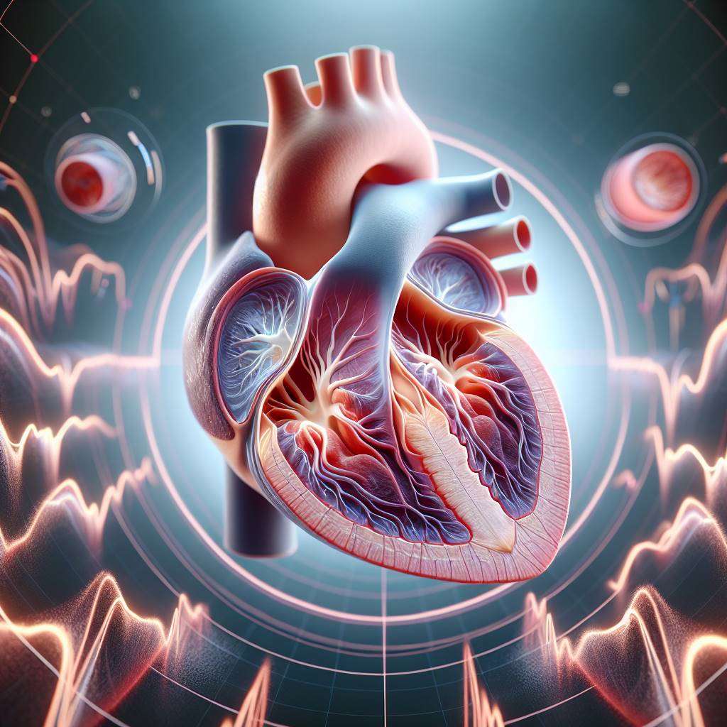
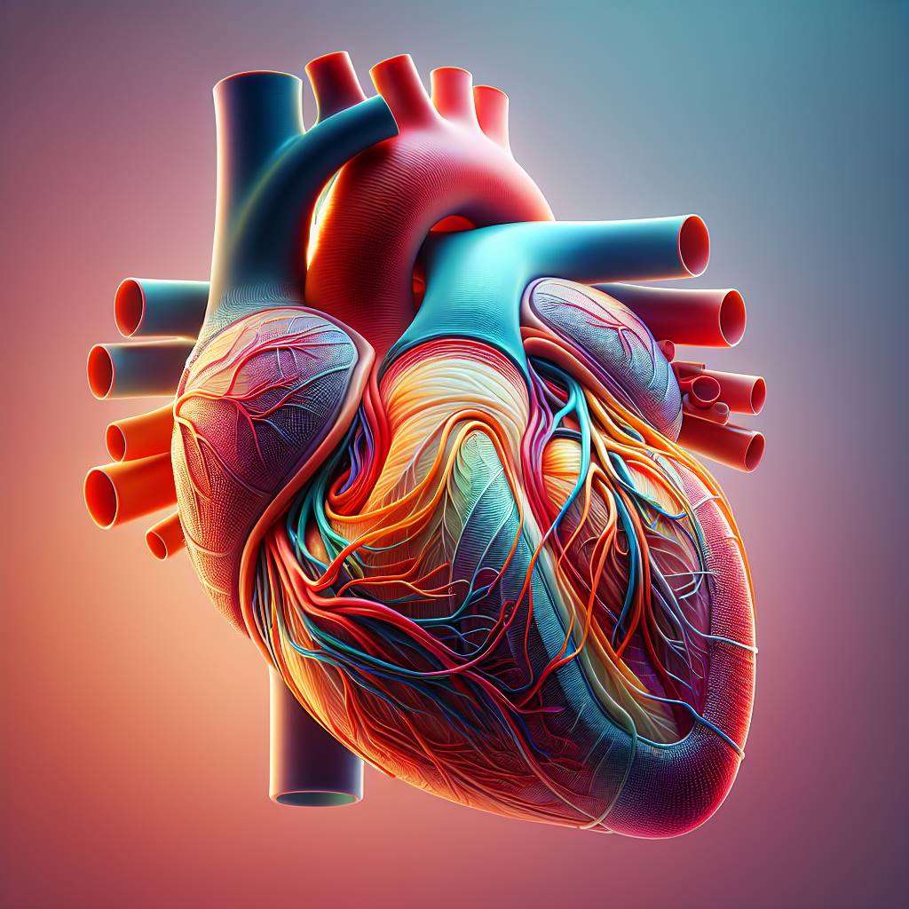
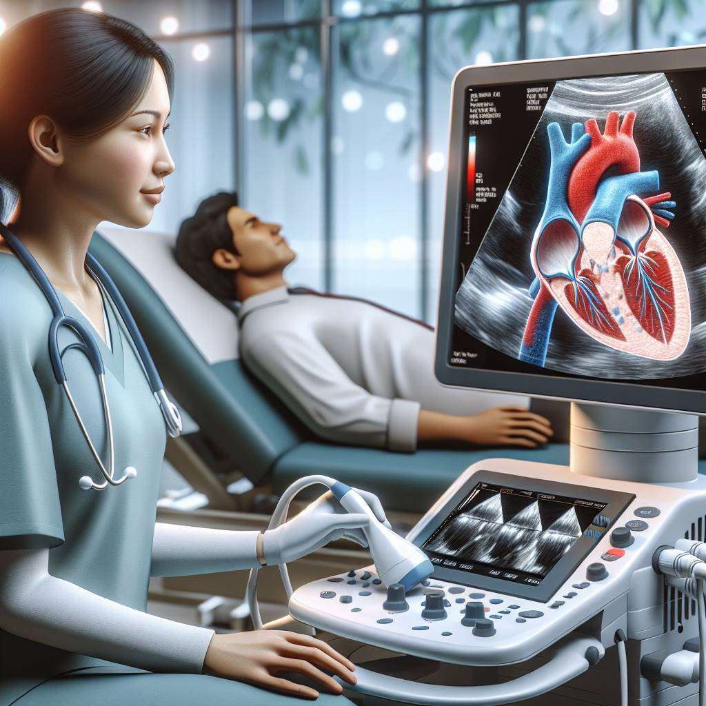
3D Echocardiography is a cutting-edge imaging technique that provides detailed, real-time views of the heart's structure and function. In cases of complex mitral valve disease, this advanced technology is invaluable for accurate diagnosis and treatment planning. It helps cardiologists visualize abnormalities in the mitral valve with unparalleled clarity, improving outcomes for patients with severe heart conditions.
Medical disclaimer: This content is for general awareness and does not replace a doctor’s consultation. For diagnosis or treatment decisions, consult a qualified specialist.
The importance of 3D echocardiography lies in its ability to guide precise interventions, especially in challenging cases of mitral regurgitation or stenosis. By offering a comprehensive view of the heart, it aids in determining the best course of treatment, whether surgical or non-invasive. This technology is transforming the management of mitral valve diseases, ensuring better care for patients.
What is 3D Echocardiography in Mitral Valve Disease?
3D echocardiography is an advanced imaging modality that uses ultrasound to create three-dimensional images of the heart. It is particularly useful in diagnosing and managing mitral valve disease, a condition where the mitral valve does not function properly. This technology allows cardiologists to assess the valve's structure and movement in real time.
Unlike traditional 2D echocardiography, 3D imaging provides a more detailed and accurate representation of the heart. This is crucial in complex cases, such as severe mitral regurgitation or stenosis, where precise visualization is necessary for effective treatment. The ability to view the heart from multiple angles helps in identifying abnormalities that might be missed with 2D imaging.
3D echocardiography is non-invasive, making it a safe and patient-friendly option. It is commonly used during pre-surgical evaluations and intraoperative procedures to ensure optimal outcomes.

How 3D Echocardiography Enhances Mitral Valve Diagnosis
The use of 3D echocardiography has revolutionized the diagnosis of mitral valve disease. This technology provides a comprehensive view of the mitral valve, enabling cardiologists to identify structural abnormalities with greater accuracy. It is particularly beneficial in detecting conditions like mitral prolapse or regurgitation, which require detailed imaging for proper assessment.
One of the key advantages of 3D echocardiography is its ability to visualize the valve's anatomy in real time. This helps in understanding the severity of the disease and planning appropriate interventions. For example, it can accurately measure the size of the valve opening, which is critical in cases of mitral stenosis.
Additionally, 3D imaging aids in detecting complications such as calcifications or chordal ruptures. This level of detail ensures that patients receive a precise diagnosis, paving the way for targeted treatments and improved outcomes.
Benefits of 3D Imaging in Complex Mitral Valve Cases
The benefits of 3D echocardiography in managing complex mitral valve disease are numerous. One of the most significant advantages is its ability to provide a detailed, three-dimensional view of the valve's structure and function. This is particularly important in cases where traditional 2D imaging falls short.
Some key benefits include:
- Enhanced visualization of the mitral valve's anatomy, including leaflets and chordae tendineae.
- Improved accuracy in diagnosing conditions like mitral regurgitation or stenosis.
- Better guidance for surgical or catheter-based interventions.
- Real-time imaging during procedures to ensure optimal outcomes.
By offering a more comprehensive view of the heart, 3D echocardiography helps in tailoring treatments to individual patient needs. This leads to better management of complex cases and improved quality of life for patients.
3D Echocardiography vs 2D: Key Differences Explained
While both 2D and 3D echocardiography are valuable tools in diagnosing mitral valve disease, there are significant differences between the two. The primary distinction lies in the level of detail and accuracy provided by 3D imaging.
The table below highlights the key differences:
| Feature |
2D Echocardiography |
3D Echocardiography |
| Imaging |
Two-dimensional, limited views |
Three-dimensional, comprehensive views |
| Accuracy |
Moderate |
High |
| Applications |
Basic diagnosis |
Complex cases and surgical planning |
These differences make 3D echocardiography the preferred choice for evaluating complex cases, ensuring better diagnostic accuracy and treatment outcomes.
Role of 3D Echocardiography in Mitral Valve Surgery
3D echocardiography plays a crucial role in the surgical management of mitral valve disease. It is widely used during preoperative planning to assess the valve's anatomy and identify any abnormalities that may impact the surgical approach. This ensures that surgeons have a clear understanding of the valve's structure before the procedure.
During surgery, 3D imaging provides real-time guidance, allowing surgeons to monitor the progress of the operation and make adjustments as needed. This is particularly important in complex cases, such as those involving mitral valve repair or replacement. The ability to visualize the valve in three dimensions ensures that the procedure is performed with precision.
Post-surgery, 3D echocardiography is used to evaluate the success of the intervention and monitor the patient's recovery. Its role in ensuring optimal surgical outcomes makes it an indispensable tool in the treatment of mitral valve disease.
Understanding Mitral Valve Anatomy Through 3D Imaging
The mitral valve plays a critical role in ensuring proper blood flow between the left atrium and left ventricle of the heart. Understanding its complex structure is essential for diagnosing and treating conditions like mitral valve stenosis and mitral regurgitation. Traditional imaging techniques often fall short in providing detailed views of the valve's anatomy.
3D echocardiography offers a revolutionary approach by creating highly detailed, real-time images of the mitral valve. This advanced imaging technique allows cardiologists to assess the valve's leaflets, annulus, and subvalvular apparatus with unparalleled precision. Such clarity is invaluable in planning surgical or interventional treatments for complex cases.
By visualizing the valve in three dimensions, clinicians can better understand abnormalities, such as prolapse or calcification, and tailor treatments to individual patients. This technology is particularly beneficial for patients with congenital or acquired heart diseases.

When is 3D Echocardiography Recommended for Mitral Valve?
3D echocardiography is recommended in several scenarios where detailed imaging of the mitral valve is crucial. It is particularly useful for evaluating patients with complex mitral valve disease, including those with severe regurgitation or stenosis. This imaging modality is also essential in pre-surgical planning, helping surgeons visualize the valve's anatomy before repair or replacement procedures.
In addition, 3D echocardiography is often used during minimally invasive procedures, such as mitral valve clip placement, to guide real-time interventions. It is also beneficial for assessing congenital abnormalities, such as cleft mitral valves, and for monitoring post-surgical outcomes to ensure the success of the treatment.
- Patients with unexplained symptoms like fatigue or shortness of breath.
- Cases where traditional 2D imaging provides inconclusive results.
- Individuals undergoing valve repair or replacement surgery.
By offering a comprehensive view of the mitral valve, 3D echocardiography ensures accurate diagnosis and effective treatment planning.
Advanced Applications of 3D Echocardiography in Cardiology
The applications of 3D echocardiography extend beyond mitral valve assessment. This cutting-edge technology is transforming the field of cardiology by enabling detailed evaluation of other heart structures, such as the aortic valve, tricuspid valve, and left atrium. It is particularly valuable in diagnosing and managing valvular heart diseases.
One of the most significant advancements is its use in guiding interventional procedures. For example, during transcatheter mitral valve repair, 3D imaging provides real-time visualization, ensuring precise device placement. It is also used in electrophysiology to map atrial fibrillation and guide catheter ablation procedures.
Moreover, 3D echocardiography is instrumental in pediatric cardiology, helping diagnose congenital heart defects with greater accuracy. Its ability to provide dynamic, real-time images makes it a powerful tool for both diagnosis and treatment in various cardiac conditions.
How Accurate is 3D Echocardiography for Valve Assessment?
The accuracy of 3D echocardiography in assessing mitral valve function is unparalleled. Studies have shown that it provides superior visualization compared to traditional 2D imaging, especially in evaluating complex conditions like mitral valve prolapse or regurgitation. Its ability to capture real-time, three-dimensional images ensures precise measurements of valve anatomy and function.
One of the key advantages of 3D echocardiography is its ability to quantify the severity of mitral regurgitation. By measuring parameters like regurgitant volume and effective regurgitant orifice area, clinicians can make more informed decisions about treatment. Additionally, it allows for accurate assessment of surgical outcomes, ensuring that repairs or replacements are functioning optimally.
| Feature |
2D Echocardiography |
3D Echocardiography |
| Image Detail |
Limited |
High |
| Real-Time Visualization |
Partial |
Comprehensive |
| Quantitative Analysis |
Basic |
Advanced |
Overall, 3D echocardiography is a game-changer in valve assessment, offering unmatched accuracy and reliability.
3D Echocardiography for Mitral Valve Regurgitation Diagnosis
Mitral valve regurgitation is a condition where the valve fails to close properly, allowing blood to flow backward into the left atrium. Diagnosing this condition accurately is critical for determining the appropriate treatment. 3D echocardiography has emerged as a gold standard for evaluating the severity and underlying causes of regurgitation.
This advanced imaging technique provides detailed views of the valve's leaflets, annulus, and subvalvular apparatus, helping clinicians identify structural abnormalities. It also allows for precise quantification of regurgitant volume and flow, which are essential for treatment planning. Furthermore, 3D echocardiography is invaluable during surgical or interventional procedures, ensuring optimal outcomes.
By offering a comprehensive and accurate assessment, 3D echocardiography plays a pivotal role in managing patients with mitral valve regurgitation, improving both diagnosis and treatment success rates.
Non-Invasive Imaging for Complex Mitral Valve Disorders
Non-invasive imaging techniques like 3D echocardiography have revolutionized the diagnosis and management of complex mitral valve disorders. This advanced imaging modality provides detailed, real-time views of the mitral valve's structure and function, aiding in accurate assessments.
Unlike traditional 2D imaging, 3D echocardiography offers a comprehensive view, enabling cardiologists to detect abnormalities such as mitral regurgitation, stenosis, or prolapse with greater precision. This is especially critical for patients with intricate valve anatomy or coexisting cardiac conditions.
Benefits of non-invasive imaging include:
- Reduced patient discomfort.
- Enhanced diagnostic accuracy.
- Improved preoperative planning.
By minimizing the need for invasive procedures, 3D echocardiography ensures safer and more effective care for patients with complex mitral valve diseases.

3D Echocardiography in Preoperative Mitral Valve Planning
Preoperative planning for mitral valve repair or replacement is critical for achieving optimal surgical outcomes. 3D echocardiography plays a pivotal role in this process by providing detailed anatomical and functional insights into the mitral valve.
Surgeons rely on this imaging technique to assess the severity of valve dysfunction, identify structural abnormalities, and determine the feasibility of repair versus replacement. The real-time imaging capabilities of 3D echocardiography allow for precise measurements of valve dimensions and leaflet motion.
Moreover, this technology helps in predicting potential complications and tailoring surgical strategies to individual patient needs. By enhancing preoperative planning, 3D echocardiography contributes to improved patient outcomes and reduced postoperative risks.
Can 3D Echocardiography Replace Traditional Imaging Methods?
The question of whether 3D echocardiography can replace traditional imaging methods like 2D echocardiography or cardiac MRI is a topic of ongoing debate. While 3D imaging offers unparalleled visualization of the mitral valve, each modality has its unique strengths.
For example, 2D echocardiography remains widely used due to its accessibility and cost-effectiveness. However, it lacks the spatial resolution and depth provided by 3D imaging. Cardiac MRI, on the other hand, excels in assessing myocardial function but is less effective for real-time valve analysis.
| Imaging Modality |
Strengths |
Limitations |
| 2D Echocardiography |
Cost-effective, widely available |
Limited spatial resolution |
| 3D Echocardiography |
Detailed valve visualization |
Higher cost, requires expertise |
| Cardiac MRI |
Excellent for myocardial assessment |
Not ideal for real-time imaging |
While 3D echocardiography is unlikely to completely replace traditional methods, it serves as a complementary tool, enhancing diagnostic accuracy and treatment planning.
Future of 3D Echocardiography in Cardiac Care
The future of 3D echocardiography in cardiac care is promising, with ongoing advancements in imaging technology and artificial intelligence (AI). These innovations aim to improve diagnostic accuracy, reduce operator dependency, and enhance patient outcomes.
AI-powered algorithms are being integrated into 3D echocardiography systems to automate image analysis and provide real-time decision support. This is particularly beneficial for detecting subtle abnormalities in mitral valve disease and other cardiac conditions.
Additionally, portable and miniaturized 3D echocardiography devices are making this technology more accessible, even in remote or resource-limited settings. As these advancements continue, 3D echocardiography is expected to play a central role in personalized cardiac care, enabling earlier diagnosis and more targeted treatments.
Challenges and Limitations of 3D Echocardiography Technology
Despite its numerous benefits, 3D echocardiography faces certain challenges and limitations. One major issue is the high cost of equipment, which can limit its availability in smaller healthcare facilities or developing regions.
Another challenge is the steep learning curve associated with interpreting 3D images. Cardiologists and technicians require specialized training to fully utilize this technology. Additionally, image quality can be affected by patient factors such as obesity or poor acoustic windows, which may hinder accurate diagnosis.
Finally, the integration of 3D echocardiography with other diagnostic tools remains a work in progress. Overcoming these limitations will require continued investment in research, training, and technology development to make this advanced imaging modality more accessible and effective.
3D Echocardiography Top Doctors in India
Dr. Naresh Trehan, Chairman and Managing Director of Medanta - The Medicity, Gurugram, is a globally renowned cardiac surgeon with over 50 years of experience. Another expert is Dr. Balbir Singh, Chairman of Cardiology at Max Super Speciality Hospital, Saket, New Delhi, with over 30 years of experience in interventional cardiology and echocardiography. Both doctors are recognized for their international expertise and innovative approaches.
Learn more on 3D Echocardiography Top Doctors in India
3D Echocardiography Top Hospitals in India
Fortis Escorts Heart Institute, New Delhi, and Apollo Hospitals, Chennai, are leading centers for advanced 3D Echocardiography. Both hospitals are NABH and JCI accredited, offering cutting-edge imaging technologies, multidisciplinary care, and robotic-assisted procedures. They also provide international patient services and have a proven track record of successful outcomes.
Find more 3D Echocardiography Top Hospitals in India
3D Echocardiography Treatment Costs in India
The cost of 3D Echocardiography in India typically ranges from ₹10,000 to ₹25,000 (approximately $120 to $300). Factors influencing costs include the hospital’s reputation, doctor’s expertise, and procedure complexity. The average hospital stay is minimal, as it is a non-invasive imaging technique. India offers a significant cost advantage compared to Western countries, with options for insurance coverage or third-party financing.
Learn 3D Echocardiography Treatment Costs in India
3D Echocardiography Treatment Procedures in India
3D Echocardiography in India is performed using advanced imaging systems to create detailed, real-time views of the heart. The procedure involves placing a transducer on the chest or inserting it through the esophagus (TEE) for enhanced visualization. Recovery is immediate as it is non-invasive. Top hospitals adopt innovative imaging technologies and adhere to global medical standards, ensuring accurate diagnosis and treatment planning.
Learn on 3D Echocardiography Treatment Procedures in India
FAQs
What is 3D Echocardiography?
3D Echocardiography is an advanced imaging technique that provides three-dimensional, real-time views of the heart, aiding in the diagnosis and management of complex cardiac conditions.
How does 3D Echocardiography help in mitral valve disease?
It offers detailed visualization of the mitral valve anatomy, helping doctors assess valve function, detect abnormalities, and plan surgical or interventional treatments with precision.
Is 3D Echocardiography safe?
Yes, it is a non-invasive and safe procedure with no known risks, making it suitable for patients of all ages, including those with critical conditions.
How long does a 3D Echocardiography procedure take?
The procedure typically takes 30 to 60 minutes, depending on the complexity of the case and the imaging requirements.
What is the difference between 2D and 3D Echocardiography?
While 2D Echocardiography provides flat, two-dimensional images, 3D Echocardiography offers detailed, three-dimensional views, enabling better visualization of cardiac structures.
Is 3D Echocardiography available in all hospitals in India?
No, it is primarily available in tertiary care centers and specialized cardiac hospitals equipped with advanced imaging technologies.
Can 3D Echocardiography replace traditional diagnostic methods?
While it enhances diagnostic accuracy, it is often used in conjunction with other tests like ECG and stress tests for comprehensive cardiac evaluation.
Is 3D Echocardiography covered by insurance in India?
Yes, most health insurance policies in India cover 3D Echocardiography if prescribed as part of a diagnostic or treatment plan.
What are the limitations of 3D Echocardiography?
It may have limitations in patients with poor acoustic windows or those unable to tolerate transesophageal imaging (TEE).
How do I prepare for a 3D Echocardiography procedure?
Preparation depends on the type of echocardiography. For transthoracic echocardiography (TTE), no special preparation is needed. For transesophageal echocardiography (TEE), fasting for a few hours may be required.
Revolutionizing Heart Health: Advances in Mitral Valve Treatment
The treatment of mitral valve conditions has seen significant advancements, particularly for patients deemed inoperable. Transcatheter Mitral Valve Replacement (TMVR) is a groundbreaking procedure that is transforming how these patients receive care. Learn more about how TMVR is changing mitral valve treatment in our detailed blog post.
Mitral valve prolapse is another critical condition that can lead to serious complications, including atrial fibrillation. Understanding the connection between these two conditions is essential for effective management and treatment. For an in-depth exploration of how mitral valve prolapse can lead to atrial fibrillation, check out our informative article.
As research and technology continue to evolve, patients with mitral valve issues have more options than ever before. Staying informed about these advancements can empower patients and healthcare providers alike to make better decisions regarding heart health.
Explore the Best Heart Care Resources in India
Find some of the top cardiologist, surgeons and the best heart hospitals in India
Best Heart Hospitals in India
Choosing the right hospital is crucial for successful heart treatments. If you want to explore trusted options, check the list of Best Heart Hospitals in India offering world-class facilities, advanced cardiac care units, and experienced teams for both simple and complex procedures.
Best Cardiologists in India
Finding the right cardiologist can make a huge difference in early diagnosis and long-term heart health. If you are looking for the Best Cardiologists in India, see this curated list of experts who specialize in preventive care, interventional cardiology, and complex heart disease management. Check the full list Best Cardiologists in India.
Best Cardiac Surgeons in India
If you are planning for heart surgery and need top-level expertise, we recommend exploring the Best Cardiac Surgeons in India. These surgeons have a proven record in performing bypass surgeries, valve replacements, and minimally invasive heart operations with excellent outcomes.
Get more indepth information on Cardiology treatments and their costs.
Conclusion
Your cardiology health deserve the best care. Explore the links above to learn more about the top cardiac hospitals and cardiac surgeons in India.