Imaging plays a vital role in the assessment of heart valve diseases, enabling healthcare professionals to accurately diagnose, evaluate, and manage various cardiac conditions. With the growing prevalence of heart valve disorders, advanced imaging techniques have become essential in understanding the structural and functional aspects of the heart valves. Proper imaging allows for better treatment planning, timely interventions, and improved patient outcomes, making it a cornerstone of modern cardiology.
Overview of Heart Valve Diseases and Their Implications
Heart valve diseases, including stenosis and regurgitation, can significantly affect the heart's ability to pump blood efficiently. These conditions may lead to symptoms such as shortness of breath, fatigue, and chest pain, and can increase the risk of serious complications like heart failure and arrhythmias. Early detection and accurate assessment of valve function are crucial for determining the appropriate treatment options, which may include medication management or surgical interventions.
Traditional Imaging Techniques for Heart Valve Assessment
Historically, several imaging modalities have been employed to assess heart valve conditions. Traditional techniques include chest X-rays, which provide a basic view of the heart's size and shape, and echocardiography, which is widely regarded as the primary method for evaluating valve function. While these traditional approaches have been beneficial, advancements in imaging technology have enhanced the ability to visualize heart valves more accurately and comprehensively.

Echocardiography: The Gold Standard in Cardiac Imaging
Echocardiography remains the gold standard for heart valve assessment due to its non-invasive nature and real-time imaging capabilities. This technique uses ultrasound waves to create detailed images of the heart's structures and can assess valve morphology, function, and blood flow patterns. Doppler echocardiography further enhances the evaluation by measuring the speed and direction of blood flow across the valves, allowing for the identification of abnormalities such as regurgitation or stenosis.
3D Echocardiography: Enhancing Visualization of Heart Valves
Recent advancements in echocardiography have introduced three-dimensional (3D) imaging, which provides a more comprehensive view of the heart valves and their surroundings. 3D echocardiography allows for detailed visualization of the valve structure, enabling clinicians to assess the severity of valve diseases more accurately. This enhanced imaging capability can aid in preoperative planning, particularly for complex cases that may require surgical intervention.
Cardiac MRI: A Non-Invasive Tool for Detailed Assessment
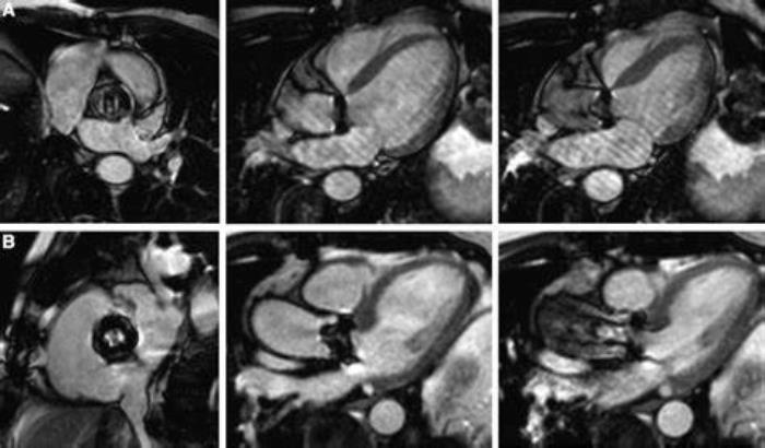
Cardiac magnetic resonance imaging (MRI) is a powerful non-invasive tool that provides high-resolution images of the heart's anatomy and function. MRI is particularly useful for assessing the heart valves, as it can provide detailed information about valve morphology and the surrounding structures. Additionally, cardiac MRI is excellent for evaluating myocardial tissue characteristics, making it a valuable resource for understanding the impact of valve disease on overall heart health.
Computed Tomography (CT) Angiography: Revolutionizing Valve Imaging
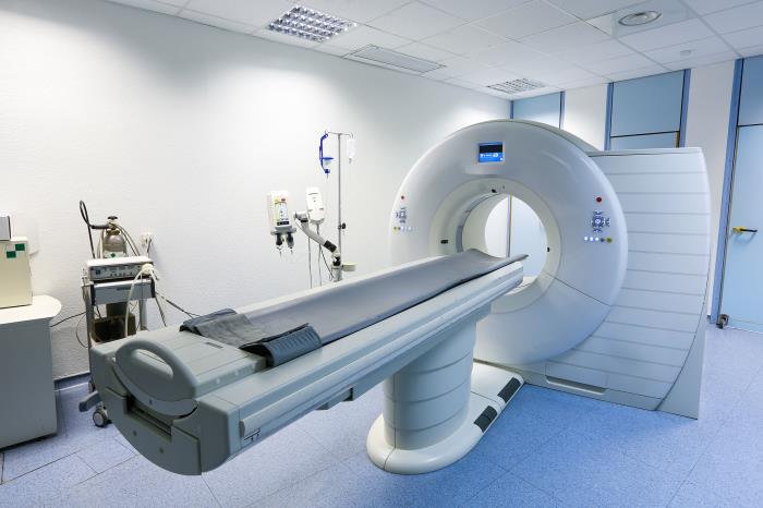
Computed tomography (CT) angiography has revolutionized the assessment of heart valves by providing detailed cross-sectional images of the heart and its blood vessels. This imaging modality is especially beneficial for evaluating complex anatomical relationships and for preoperative planning in patients undergoing valve surgery. CT angiography can also help identify coronary artery disease, which is crucial for comprehensive cardiac assessments, ensuring that patients receive the most appropriate treatment for their heart valve conditions.

The Role of Transesophageal Echocardiography (TEE)
Transesophageal echocardiography (TEE) is a specialized imaging technique used to obtain detailed views of the heart's structures, including the heart valves, chambers, and blood vessels. Unlike transthoracic echocardiography (TTE), which uses external probes, TEE involves inserting a probe into the esophagus, allowing for clearer and more accurate images. This technique is particularly useful for assessing complex valve disorders, guiding surgical interventions, and monitoring patients postoperatively. TEE provides critical information that enhances the accuracy of diagnoses and the effectiveness of treatment plans.
Innovations in Real-Time Imaging Techniques
Advancements in real-time imaging techniques have significantly improved the assessment of heart valves and overall cardiac function. Technologies such as 3D echocardiography allow clinicians to visualize the heart in three dimensions, providing a comprehensive view of valve morphology and function. Innovations in ultrasound technology have also led to enhanced resolution and faster image acquisition, enabling clinicians to make timely and informed decisions during surgical procedures. Real-time imaging improves diagnostic capabilities and allows for immediate adjustments during interventions, thereby enhancing patient outcomes.
Artificial Intelligence in Imaging: Enhancing Accuracy and Efficiency
The integration of artificial intelligence (AI) in cardiac imaging is transforming how heart conditions, including valve diseases, are diagnosed and managed. AI algorithms can analyze imaging data quickly and accurately, identifying patterns that may be missed by the human eye. These tools enhance diagnostic accuracy and efficiency, allowing healthcare professionals to focus on patient care. AI can also assist in automating routine tasks, reducing the time required for image analysis and improving workflow in clinical settings. As AI continues to evolve, its role in heart valve imaging is expected to expand, leading to better patient outcomes.
Advanced Doppler Imaging: Assessing Blood Flow Dynamics
Advanced Doppler imaging techniques play a crucial role in evaluating blood flow dynamics across heart valves. By measuring the velocity of blood flow, Doppler imaging can assess the severity of valve stenosis or regurgitation, providing valuable information for treatment decisions. Techniques such as color Doppler and spectral Doppler allow for the visualization of blood flow patterns and help clinicians quantify the degree of hemodynamic impact caused by valve abnormalities. This information is essential for planning interventions and monitoring postoperative outcomes.
Hybrid Imaging Techniques: Combining Modalities for Comprehensive Assessment
Hybrid imaging techniques combine multiple imaging modalities to provide a comprehensive assessment of heart valves and overall cardiac health. For example, integrating echocardiography with computed tomography (CT) or magnetic resonance imaging (MRI) allows for detailed anatomical visualization while simultaneously assessing functional parameters. This multifaceted approach enables clinicians to develop personalized treatment plans tailored to the individual patient's needs, improving diagnostic accuracy and surgical outcomes.
The Impact of Imaging on Surgical Planning and Decision-Making
Imaging plays a pivotal role in surgical planning and decision-making for heart valve procedures. Detailed imaging studies help identify the specific anatomy of the heart valves, assess the severity of any underlying conditions, and plan the optimal surgical approach. Surgeons can visualize the extent of calcification, determine the appropriate type of replacement valve, and anticipate potential complications during surgery. Accurate imaging enhances the surgeon's ability to perform complex procedures effectively, ultimately leading to improved patient outcomes.
Preoperative Imaging: Identifying Risks and Complications
Preoperative imaging is essential for identifying potential risks and complications before heart valve surgery. Imaging studies can reveal underlying conditions such as pulmonary hypertension, left ventricular dysfunction, or aortic disease, which may affect surgical outcomes. By identifying these risks in advance, healthcare providers can implement strategies to mitigate them, ensuring that patients are better prepared for surgery. This proactive approach enhances the safety and effectiveness of surgical interventions.
Postoperative Imaging: Monitoring Valve Function and Complications
Postoperative imaging is crucial for monitoring heart valve function and detecting any complications after surgery. Regular imaging assessments, including echocardiography and other modalities, allow healthcare providers to evaluate the effectiveness of the intervention and identify issues such as valve dysfunction or thrombus formation. Early detection of complications can lead to timely interventions, improving patient prognosis and quality of life following valve surgery.
Patient Safety in Imaging: Minimizing Risks and Radiation Exposure
Ensuring patient safety during imaging procedures is of utmost importance. While many imaging techniques, such as echocardiography, carry minimal risks, others, like CT scans, involve exposure to radiation. Healthcare providers must weigh the benefits of advanced imaging against the potential risks and employ strategies to minimize exposure. This includes using the lowest effective radiation dose, optimizing imaging protocols, and considering alternative imaging modalities when appropriate. Educating patients about the safety measures in place can help alleviate their concerns.
Cost-Effectiveness of Advanced Imaging Technologies
The cost-effectiveness of advanced imaging technologies is a significant consideration in modern healthcare. While these technologies may involve higher initial costs, their ability to improve diagnostic accuracy, reduce unnecessary procedures, and enhance patient outcomes can lead to overall cost savings. Studies have shown that effective imaging can shorten hospital stays, decrease the need for repeat interventions, and improve long-term health outcomes, ultimately justifying the investment in advanced imaging technologies.
Future Directions in Heart Valve Imaging Technology
The future of heart valve imaging technology is promising, with ongoing research and innovation expected to yield new advancements. Emerging technologies, such as molecular imaging and portable imaging devices, may further enhance the ability to diagnose and monitor heart valve diseases. Additionally, advancements in AI and machine learning are anticipated to improve image interpretation and automate routine tasks, freeing healthcare providers to focus on patient care. As these technologies evolve, they will play an increasingly important role in improving heart valve care.
Case Studies: Successful Outcomes Through Advanced Imaging
Numerous case studies demonstrate the positive impact of advanced imaging technologies on patient outcomes in heart valve care. For example, patients who underwent preoperative 3D echocardiography prior to valve replacement surgery experienced fewer complications and shorter recovery times. Similarly, cases where hybrid imaging techniques were utilized allowed for more precise surgical planning, leading to improved postoperative results. These success stories highlight the importance of advanced imaging in achieving optimal outcomes for patients with heart valve disorders.
Understanding Anticoagulation Therapy Post Heart Valve Replacement
Learn about anticoagulation therapy after heart valve replacement. This section discusses the importance of blood-thinning medications, their role in preventing blood clots, and the necessary monitoring and lifestyle adjustments patients must consider to ensure their safety and well-being.
Innovations in Heart Valve Surgery: Minimally Invasive Techniques
Stay informed about the innovations in heart valve surgery, particularly minimally invasive techniques. This section explores how these advancements reduce recovery time, minimize scarring, and improve patient outcomes, revolutionizing the way heart valve surgeries are performed.
Conclusion: The Role of Advanced Imaging in Improving Heart Valve Care
Advanced imaging technologies are integral to the assessment and management of heart valve diseases. From initial diagnosis to surgical planning and postoperative monitoring, imaging plays a critical role in ensuring optimal patient outcomes. As technology continues to advance, the potential for improved accuracy, efficiency, and safety in heart valve care will only increase. By embracing these innovations, healthcare providers can enhance their ability to deliver high-quality care to patients with heart valve disorders.
Best Heart Valve Replacement Surgeons in India
The Best Heart Valve Replacement Surgeons in India are highly skilled in complex valve procedures, offering personalized care to help patients achieve successful outcomes and enhanced heart health.
FAQ
What are the most common imaging techniques used for heart valve assessment?
Common imaging techniques include transthoracic echocardiography (TTE), transesophageal echocardiography (TEE), cardiac MRI, and Doppler imaging.
How does 3D echocardiography improve valve visualization?
3D echocardiography provides detailed, volumetric images of heart structures, allowing for better visualization of valve anatomy and function compared to traditional 2D imaging.
What role does cardiac MRI play in evaluating heart valves?
Cardiac MRI provides high-resolution images and can assess both the structure and function of heart valves, as well as evaluate associated conditions like myocardial infarction.
How has artificial intelligence impacted imaging for heart valve assessment?
AI enhances imaging by improving diagnostic accuracy through pattern recognition, automating routine analyses, and aiding in treatment planning, thus streamlining workflow.
What should patients expect during the imaging process for heart valve evaluation?
Patients can expect to undergo a non-invasive procedure involving sound waves (for echocardiography) or a brief MRI scan, during which they may need to lie still and follow specific instructions.
Explore the Best Heart Care Resources in India
Find some of the top cardiologist, surgeons and the best heart hospitals in India
Best Heart Hospitals in India
Choosing the right hospital is crucial for successful heart treatments. If you want to explore trusted options, check the list of Best Heart Hospitals in India offering world-class facilities, advanced cardiac care units, and experienced teams for both simple and complex procedures.
Best Cardiologists in India
Finding the right cardiologist can make a huge difference in early diagnosis and long-term heart health. If you are looking for the Best Cardiologists in India, see this curated list of experts who specialize in preventive care, interventional cardiology, and complex heart disease management. Check the full list Best Cardiologists in India.
Best Cardiac Surgeons in India
If you are planning for heart surgery and need top-level expertise, we recommend exploring the Best Cardiac Surgeons in India. These surgeons have a proven record in performing bypass surgeries, valve replacements, and minimally invasive heart operations with excellent outcomes.