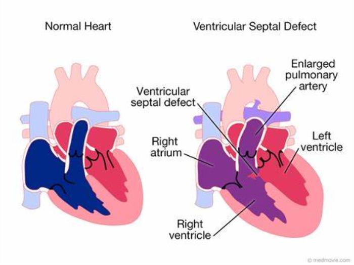
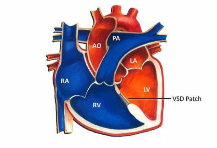
A Ventricular Septal Defect (VSD) is a congenital heart defect characterized by an abnormal opening between the left and right ventricles of the heart. This condition allows oxygen-rich blood from the left ventricle to mix with oxygen-poor blood in the right ventricle, which can lead to increased workload on the heart and lungs. VSDs can vary in size and severity, and some may cause significant symptoms, while others may remain asymptomatic for years.
The Importance of Cardiac Imaging in Diagnosing VSD
Cardiac imaging plays a vital role in the diagnosis of Ventricular Septal Defect (VSD). Since VSDs can range from small, inconsequential holes to large defects that significantly affect heart function, accurate imaging is essential in understanding the size, location, and impact of the defect. This information helps cardiologists plan the best course of treatment, whether it involves monitoring, medication, or surgery.
Common Imaging Techniques Used to Diagnose VSD
Several imaging techniques are used to diagnose Ventricular Septal Defect (VSD), each offering unique benefits. The primary goal is to visualize the heart's structures and detect the presence of a septal defect. Among the most commonly used techniques are echocardiography, MRI, CT scans, and X-rays. Each technique provides valuable insights that guide the management of the condition, with some methods being more suitable for specific patient cases.

Echocardiography: The Gold Standard for VSD Diagnosis
Echocardiography is the gold standard for diagnosing Ventricular Septal Defect (VSD) because it is non-invasive, widely available, and provides real-time images of the heart. This technique uses sound waves to produce detailed images of the heart's structures and blood flow. It is especially effective in detecting the size and location of the VSD, as well as assessing the impact of the defect on heart function. Doppler echocardiography can also evaluate the flow of blood through the VSD, helping to determine the severity of the defect and its potential impact on the patient’s health. In most cases, echocardiography is the first imaging method used to confirm a VSD diagnosis.
Cardiac MRI in VSD Diagnosis: When Is It Used?
Cardiac MRI is an advanced imaging technique used in Ventricular Septal Defect (VSD) diagnosis, particularly in complex cases or when echocardiographic results are inconclusive. MRI provides high-resolution images and detailed information about the heart's structure and function. It is especially useful in evaluating the size and location of a VSD, especially when the defect is located in areas that are difficult to assess with other imaging methods. Additionally, MRI can assess heart muscle function and detect any complications that may result from a VSD, such as right or left ventricular enlargement. While more expensive and less widely available than echocardiography, MRI is an important tool in cases where more detailed information is needed.
CT Scans for VSD: A Useful Tool in Complex Cases
CT scans (computed tomography) are occasionally used for the diagnosis of Ventricular Septal Defect (VSD), particularly in complex or challenging cases where other imaging techniques may not provide sufficient information. CT scans can offer detailed, cross-sectional images of the heart and can help visualize the anatomy of the VSD and its surrounding structures. This technique is especially helpful for planning surgical interventions or assessing the anatomy of patients with complex congenital heart defects. However, CT scans involve exposure to radiation and are generally used when other non-invasive methods like echocardiography and MRI are insufficient.
Role of X-ray Imaging in the Diagnosis of VSD
X-ray imaging can play a supportive role in diagnosing Ventricular Septal Defect (VSD), especially in the context of detecting related complications. While X-rays cannot directly visualize the VSD, they can reveal signs of heart enlargement or pulmonary congestion, which may be indicative of a significant VSD. An X-ray is often used to assess the overall size and shape of the heart, which can be useful in cases where VSDs cause a noticeable enlargement of the heart chambers or complications such as fluid buildup in the lungs (pulmonary edema). However, X-ray imaging is typically not used as a primary diagnostic tool for VSD and is often complemented by more advanced imaging techniques like echocardiography or MRI.
How Cardiac Imaging Helps Determine VSD Size and Location
Cardiac imaging is essential for accurately determining the size and location of a ventricular septal defect (VSD). Techniques such as echocardiography, MRI, and CT scans provide detailed views of the heart’s structure, allowing physicians to measure the size of the VSD and pinpoint its exact location within the ventricular septum. This information is critical for selecting the most appropriate treatment, whether it involves surgical repair, catheter-based procedures, or medical management.
The Role of Cardiac Imaging in Assessing VSD Severity
Cardiac imaging plays a vital role in assessing the severity of a VSD by visualizing the size of the hole, the direction and volume of blood flow, and the degree of pressure or strain on the heart’s chambers. Severe VSDs can cause significant shunting of blood between the ventricles, leading to increased pulmonary circulation and potential heart failure. Imaging helps classify VSD severity, which is crucial for deciding whether surgical intervention is necessary and for planning the type of surgery needed.
3D Echocardiography for Precise VSD Diagnosis and Surgical Planning
3D echocardiography provides detailed, real-time, three-dimensional images of the heart, offering a clearer and more accurate view of the VSD than traditional 2D imaging. This advanced imaging technique allows surgeons to assess the size, shape, and location of the VSD in a more comprehensive way, which is invaluable for pre-surgical planning. It also helps in evaluating the surrounding structures, such as valves and other heart chambers, ensuring a precise repair and reducing the risk of complications.
Advancements in Imaging Technology for More Accurate VSD Detection
Recent advancements in cardiac imaging, such as higher-resolution echocardiography, MRI, and CT scans, have greatly improved the accuracy of VSD detection. These technologies allow for better visualization of complex VSDs that may have previously been difficult to detect. High-resolution imaging provides more detailed anatomical information, allowing doctors to detect even small defects, monitor blood flow, and assess the function of the heart, improving diagnostic accuracy and patient outcomes.

How Cardiac Imaging Aids in Pre-Surgery Planning for VSD Repair
Cardiac imaging is instrumental in pre-surgery planning for VSD repair. By providing detailed views of the VSD, its surrounding structures, and the heart’s overall function, imaging allows surgeons to plan the most effective approach for repair. For example, imaging can identify whether a VSD is located near critical structures such as the aortic valve, helping the surgeon avoid damage during the procedure. Imaging also helps in selecting the best surgical technique, whether through open surgery or minimally invasive methods.
Imaging in Post-Surgery VSD Monitoring: What to Expect
After VSD surgery, cardiac imaging is used to monitor the success of the repair and ensure that the defect is fully closed. Follow-up imaging, such as echocardiography, helps detect any residual shunting or complications, such as valve dysfunction, that might arise after surgery. Regular imaging is essential to track the heart’s function and assess for any changes that could require further intervention.
The Impact of Cardiac Imaging on Minimizing Surgical Risks in VSD Treatment
Cardiac imaging helps minimize surgical risks by providing surgeons with detailed information about the defect’s size, location, and potential complications. Pre-operative imaging ensures that surgeons can plan for the best approach, reducing the likelihood of complications such as injury to surrounding tissues or improper closure of the VSD. Real-time imaging during surgery also allows for immediate adjustments, ensuring a safer procedure and more favorable outcomes.
Imaging-Based Decision Making: Choosing the Right VSD Treatment Approach
Imaging plays a key role in deciding the most appropriate treatment for a VSD. Based on imaging results, physicians can determine whether a defect can be treated with catheter-based closure, minimally invasive surgery, or traditional open-heart surgery. For larger or more complex defects, surgery may be necessary, whereas smaller or less symptomatic VSDs might be monitored or treated non-surgically. Imaging provides the evidence needed to make informed decisions about treatment options, improving the chances of successful outcomes.
The Role of Imaging in Identifying VSD-Related Complications
Cardiac imaging is vital in identifying complications associated with VSDs, such as pulmonary hypertension, heart failure, or damage to other cardiac structures. Imaging can reveal changes in blood flow patterns, ventricular size, or valve function that indicate the presence of complications. Early detection of these issues allows for timely intervention, potentially preventing more serious conditions such as arrhythmias or further cardiac damage.
Non-Invasive Imaging Techniques for Follow-Up Care After VSD Repair
Non-invasive imaging techniques, primarily echocardiography, are commonly used in follow-up care after VSD repair. These techniques are effective in monitoring the heart’s structure and function without the need for invasive procedures. Regular follow-up with echocardiograms can help ensure that the VSD repair remains intact and that the heart continues to function well. Non-invasive imaging also helps detect any late complications, such as the development of new defects or issues with heart valves.
The Future of Cardiac Imaging in VSD Diagnosis and Treatment
The future of cardiac imaging in VSD diagnosis and treatment is promising, with advances in imaging technology that promise to provide even more detailed and accurate information. For example, improvements in MRI and CT scanning can provide 3D images with higher resolution, allowing for better assessment of complex VSDs. The continued development of non-invasive imaging techniques will likely improve patient comfort, reduce risks, and enhance post-operative monitoring.
How Cardiac Imaging Improves Outcomes for VSD Patients
Cardiac imaging improves outcomes for VSD patients by ensuring accurate diagnosis, precise surgical planning, and careful post-operative monitoring. By providing real-time data, imaging helps doctors make timely and informed decisions, leading to better treatment outcomes. With advancements in imaging technologies, VSD patients can benefit from more effective and personalized care, reducing complications and improving quality of life.
Conclusion: The Critical Role of Imaging in Comprehensive VSD Care
Cardiac imaging plays a critical role throughout the care continuum for VSD patients—from diagnosis and pre-surgery planning to post-surgery monitoring and long-term follow-up. With its ability to provide detailed, accurate information, imaging ensures that healthcare providers can make informed decisions, minimize risks, and achieve the best possible outcomes for VSD patients. As imaging technology continues to evolve, it will further enhance the precision and effectiveness of VSD treatment.
Best VSD Surgery in India
The Best VSD Surgery in India provides a highly effective treatment for patients with a hole in the heart's ventricular wall, improving heart function and preventing complications.
Best VSD Surgery Hospitals in India
The best vsd surgery hospitals in india offer state-of-the-art medical facilities and expert pediatric cardiologists to ensure the best care for patients undergoing VSD repair surgery.
VSD Surgery Cost in India
The vsd surgery cost in india is affordable, with transparent pricing that provides high-quality care options for those in need of heart defect treatment.
Best VSD Surgery Surgeons in India
The Best VSD Surgery Surgeons in India are highly skilled in heart defect repairs, offering personalized care and advanced surgical techniques for successful outcomes.
FAQ
What imaging techniques are used to diagnose VSD?
Echocardiography is the most commonly used technique for diagnosing VSD, but CT and MRI may also be used for more detailed assessments, particularly for complex cases.
How does echocardiography help in diagnosing VSD?
Echocardiography uses sound waves to create images of the heart, helping to visualize the VSD’s size, location, and the impact on blood flow. It is a non-invasive, real-time technique commonly used in VSD diagnosis.
When is MRI or CT used for VSD diagnosis?
MRI or CT is typically used when echocardiography does not provide sufficient detail or when the VSD is complex. These imaging techniques provide a more comprehensive view of the heart’s structures and can be helpful in pre-surgical planning.
How does imaging help in planning surgery for VSD?
Imaging allows surgeons to assess the VSD’s size, location, and any potential complications, helping them plan the most appropriate surgical approach to repair the defect effectively and safely.
What role does cardiac imaging play in post-surgery monitoring for VSD?
Post-surgery, imaging is used to ensure that the VSD repair is intact and monitor the heart’s function. It helps detect any residual defects, complications, or changes in blood flow that may require further treatment or intervention.