Imaging plays a critical role in angioplasty, providing detailed visualization of the coronary arteries and allowing for precise assessment and treatment of blockages. By utilizing advanced imaging techniques, physicians can accurately identify the location and severity of arterial blockages, select appropriate intervention methods, and ensure optimal placement of stents. Effective imaging not only improves procedural success but also helps reduce the risk of complications, contributing to safer outcomes for patients.
What is Angioplasty? A Quick Overview
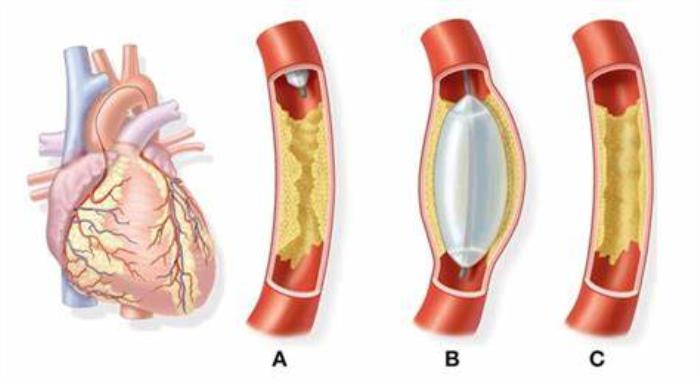
Angioplasty is a minimally invasive procedure performed to restore blood flow in blocked or narrowed coronary arteries. During the procedure, a catheter with a small balloon is guided to the blockage site. The balloon is then inflated to compress plaque against the artery walls, widening the artery. In many cases, a stent—a small, mesh-like tube—is inserted to help keep the artery open. Angioplasty is commonly used to alleviate symptoms of coronary artery disease, such as chest pain and shortness of breath, and to prevent heart attacks.
Why Imaging is Essential in Angioplasty Procedures
Imaging is essential in angioplasty because it provides real-time visualization of the arteries, enabling physicians to make informed decisions during the procedure. Precise imaging allows for accurate identification of blockages, aids in the correct placement of catheters and stents, and ensures that the blood vessels are adequately widened. Additionally, imaging helps monitor the procedure’s progress and assess potential complications, leading to safer and more effective outcomes for patients.

Types of Imaging Techniques Used in Angioplasty
Various imaging techniques are utilized in angioplasty to enhance procedural accuracy and safety. Traditional X-ray angiography is widely used to obtain clear images of blood vessels, allowing clinicians to navigate and treat blockages. Intravascular ultrasound (IVUS) and optical coherence tomography (OCT) are also employed to provide high-resolution images from within the artery itself, offering detailed insights into plaque composition and arterial structure. These advanced techniques aid in refining treatment approaches and improving the overall success of angioplasty procedures.
X-Ray Angiography: The Traditional Standard
X-ray angiography has been the standard imaging technique in angioplasty for many years. It involves injecting a contrast dye into the arteries to make them visible on X-ray images, allowing doctors to identify and target blockages with precision. Although effective, X-ray angiography provides a two-dimensional view, which can sometimes limit the amount of information available about the plaque structure and arterial walls. Despite this limitation, it remains a valuable tool in guiding angioplasty and assessing the immediate outcome of the procedure.
Intravascular Ultrasound (IVUS) and Its Benefits
Intravascular ultrasound (IVUS) is an advanced imaging technique that provides detailed, real-time images of the artery from within. By using sound waves to visualize the artery walls and plaque buildup, IVUS offers a more comprehensive view than traditional X-ray angiography. This enables physicians to assess the size and composition of plaque, optimize stent placement, and ensure effective dilation of the artery. IVUS is particularly beneficial for complex cases where precise imaging is crucial for achieving optimal outcomes.
Optical Coherence Tomography (OCT) in Detailed Artery Imaging
Optical Coherence Tomography (OCT) is a valuable imaging tool in angioplasty, allowing detailed visualization of arterial walls and blockages. OCT provides high-resolution, cross-sectional images, which help doctors assess the condition of arteries and make precise decisions on stent placement.
The Role of Fractional Flow Reserve (FFR) in Assessing Blockages
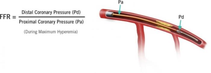
Fractional Flow Reserve (FFR) is a technique used to measure blood pressure differences across coronary artery blockages. FFR helps determine the severity of blockages and whether they require intervention, optimizing treatment plans and improving patient outcomes.

Comparing IVUS and OCT: Which is Better?
Intravascular Ultrasound (IVUS) and OCT are both powerful tools in angioplasty. IVUS offers deeper penetration, suitable for larger and complex blockages, while OCT provides greater resolution, making it ideal for detecting small details in arterial walls. Choosing between the two depends on the specific needs of each case.
How Imaging Improves Precision in Stent Placement
Advanced imaging techniques enable precise stent placement by visualizing the exact location and dimensions of arterial blockages. This precision minimizes the risk of complications and improves long-term outcomes, as stents are positioned accurately to maintain optimal blood flow.
Advanced 3D Imaging and Its Impact on Angioplasty
3D imaging advancements have revolutionized angioplasty by providing detailed, multi-dimensional views of arteries. These views aid in identifying blockages and assist in planning the procedure, ultimately improving patient safety and reducing complications.

Reducing Radiation Exposure Through Improved Imaging
Innovations in imaging technology focus on reducing radiation exposure during angioplasty. Techniques such as low-dose fluoroscopy and real-time 3D imaging enable high-quality images with minimal radiation, enhancing patient safety without compromising precision.
Imaging in Real-Time: Enhancing Patient Safety and Outcomes
Real-time imaging allows surgeons to make on-the-spot adjustments during angioplasty, enhancing both safety and effectiveness. This technology ensures that blockages are adequately treated, which improves overall patient outcomes.
The Use of Non-Invasive Imaging Before Angioplasty
Non-invasive imaging, like CT angiography, is often used before angioplasty to assess arterial blockages and plan treatment. These methods provide valuable insights while avoiding the risks associated with invasive procedures, making them an important step in pre-procedure planning.
Patient Preparation for Imaging During Angioplasty
Preparation for imaging includes instructions on fasting, hydration, and medication adjustments. Clear communication about the imaging process can help patients feel more comfortable and ensures the procedure runs smoothly.
Risks and Limitations of Imaging Techniques
Although imaging techniques like OCT and IVUS are highly effective, they do carry some risks, including contrast dye reactions and rare cases of vessel injury. Limitations also exist, such as the inability of OCT to penetrate deeper tissues, which may necessitate complementary imaging methods.
Technological Advancements in Imaging for Angioplasty
Technological advancements continue to improve the accuracy, speed, and safety of imaging in angioplasty. Innovations such as AI-driven imaging and enhanced 3D reconstruction promise to further refine angioplasty procedures, benefiting both patients and medical professionals.
How Imaging Impacts Long-Term Angioplasty Outcomes
Accurate imaging during angioplasty has a direct impact on long-term outcomes by ensuring proper stent placement and monitoring for potential complications. High-quality imaging reduces the likelihood of re-narrowing and improves overall heart health.
Future Directions: Innovations in Imaging for Angioplasty
Future advancements in imaging are likely to focus on improving resolution, reducing risks, and increasing non-invasive options. Emerging technologies, such as AI and machine learning, are expected to play a significant role in enhancing imaging precision and treatment planning.
Angioplasty and Diabetes: What Patients Need to Know
Discover critical information about angioplasty in the context of diabetes. This section discusses the unique considerations and risks for diabetic patients, emphasizing the importance of effective blood sugar management and collaboration with healthcare providers to ensure successful surgery and recovery.
Exploring the Latest Research in Coronary Angioplasty
Stay updated with the latest advancements in coronary angioplasty research. This section reviews recent studies, emerging technologies, and evolving practices that are shaping the future of angioplasty, aiming to improve patient outcomes and procedural success rates.
Conclusion: The Evolving Role of Imaging in Successful Angioplasty
Imaging continues to evolve as a fundamental component of successful angioplasty, enhancing precision, safety, and patient outcomes. As technology progresses, imaging techniques will become even more integral in guiding treatment decisions and optimizing patient care.
Best Coronary Angioplasty in India
The Best Coronary Angioplasty in India is a minimally invasive procedure that helps restore blood flow to the heart by widening blocked arteries, improving cardiovascular health and reducing chest pain.
Best Coronary Angioplasty Hospitals in India
The best coronary angioplasty hospitals in india offer state-of-the-art facilities and highly skilled cardiology teams, ensuring comprehensive pre- and post-operative care for effective treatment outcomes.
Coronary Angioplasty Cost in India
The coronary angioplasty cost in india is affordable, with transparent pricing and flexible options, making high-quality cardiovascular care accessible for patients.
Best Coronary Angioplasty Surgeons in India
The Best Coronary Angioplasty Surgeons in India are experienced specialists in interventional cardiology, dedicated to providing personalized and effective treatment for each patient.
FAQ
What imaging techniques are commonly used in angioplasty?
Common techniques include Optical Coherence Tomography (OCT), Intravascular Ultrasound (IVUS), and Fractional Flow Reserve (FFR), each providing unique insights for procedure planning and stent placement.
How does imaging improve the accuracy of angioplasty procedures?
Imaging provides detailed views of blockages and artery walls, enabling precise stent placement and reducing the risk of complications, which enhances the overall success of the procedure.
What is the difference between IVUS and OCT in angioplasty?
IVUS offers deeper penetration for visualizing larger structures, while OCT provides higher resolution, better suited for identifying smaller details in the arteries.
Are there risks associated with imaging during angioplasty?
Yes, there are minimal risks, such as contrast dye reactions and rare instances of vessel injury, but these are outweighed by the benefits of accurate imaging.
How does imaging affect the success rate of angioplasty?
High-quality imaging improves success rates by enabling accurate stent placement, reducing complications, and supporting better long-term outcomes for patients.
Explore the Best Heart Care Resources in India
Find some of the top cardiologist, surgeons and the best heart hospitals in India
Best Heart Hospitals in India
Choosing the right hospital is crucial for successful heart treatments. If you want to explore trusted options, check the list of Best Heart Hospitals in India offering world-class facilities, advanced cardiac care units, and experienced teams for both simple and complex procedures.
Best Cardiologists in India
Finding the right cardiologist can make a huge difference in early diagnosis and long-term heart health. If you are looking for the Best Cardiologists in India, see this curated list of experts who specialize in preventive care, interventional cardiology, and complex heart disease management. Check the full list Best Cardiologists in India.
Best Cardiac Surgeons in India
If you are planning for heart surgery and need top-level expertise, we recommend exploring the Best Cardiac Surgeons in India. These surgeons have a proven record in performing bypass surgeries, valve replacements, and minimally invasive heart operations with excellent outcomes.