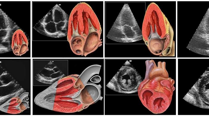
Echocardiography plays a pivotal role in diagnosing and monitoring mitral valve disease, offering detailed, non-invasive imaging of the heart's structures and functions. This diagnostic tool allows healthcare providers to assess the condition of the mitral valve, detect abnormalities, and determine the appropriate course of treatment. Through the use of high-frequency sound waves, echocardiography provides real-time images that help identify issues such as mitral valve prolapse, stenosis, or regurgitation, all of which can significantly affect heart function.
What is Echocardiography? Understanding the Basics of the Procedure
Echocardiography is an imaging technique that uses ultrasound waves to create detailed pictures of the heart’s chambers, valves, and blood flow. During the procedure, a device called a transducer emits sound waves, which bounce off heart structures and return to the device, where they are converted into visual images. This method is safe, non-invasive, and does not involve radiation, making it an essential tool for diagnosing heart conditions, including mitral valve disease. The resulting images help doctors assess the heart's anatomy, function, and blood flow, providing critical information for diagnosis and treatment.
Types of Echocardiograms Used to Diagnose Mitral Valve Disease
There are several types of echocardiograms that can be used to diagnose mitral valve disease, each providing different levels of detail and insight into the heart’s structure. The two most commonly used types are transthoracic echocardiograms (TTE) and transesophageal echocardiograms (TEE). TTE is the standard method, often used as a first-line diagnostic tool, while TEE provides more detailed images and is typically used when a clearer view of the heart’s internal structures is needed.
Transthoracic Echocardiogram (TTE): The Standard Diagnostic Tool
The transthoracic echocardiogram (TTE) is the most commonly used echocardiographic test for diagnosing mitral valve disease. In this procedure, a technician places a gel on the patient’s chest and moves a small device, known as a transducer, over the skin. The transducer emits sound waves that bounce off the heart, creating images that are displayed on a monitor. TTE is a non-invasive, straightforward procedure that provides valuable information about the mitral valve's function and structure, making it a critical tool in diagnosing mitral valve prolapse, stenosis, and regurgitation.

Transesophageal Echocardiogram (TEE): A More Detailed View of the Heart
While the transthoracic echocardiogram is the initial test of choice, a transesophageal echocardiogram (TEE) can be used when a more detailed view is required. In this procedure, a specialized probe is inserted into the patient’s esophagus, which lies closer to the heart, allowing for clearer images. TEE is particularly helpful in identifying mitral valve abnormalities, such as mitral valve prolapse or regurgitation, that may be difficult to detect with a standard TTE. TEE is often performed when the TTE results are inconclusive or when further assessment is necessary for surgical planning or valve repair.
How Echocardiography Helps Identify Mitral Valve Prolapse and Stenosis
Echocardiography is crucial in diagnosing two common forms of mitral valve disease: mitral valve prolapse (MVP) and mitral stenosis. MVP occurs when the valve’s leaflets bulge into the left atrium, which can lead to valve leakage (regurgitation). Echocardiograms can clearly show the movement of the mitral valve leaflets and reveal whether they are prolapsing. Mitral stenosis, on the other hand, involves the narrowing of the valve opening, which can impede blood flow. Echocardiography allows doctors to measure the size of the valve opening and assess blood flow patterns, helping to diagnose stenosis and determine its severity.
Role of Echocardiography in Assessing Mitral Valve Regurgitation
Mitral valve regurgitation occurs when the mitral valve doesn’t close properly, allowing blood to flow backward into the left atrium. This condition can lead to heart failure if left untreated. Echocardiography plays a crucial role in assessing the severity of mitral regurgitation by visualizing the amount of blood leaking back through the valve and evaluating the size and function of the left atrium and ventricle. Doppler imaging, a feature of echocardiography, can measure the speed and direction of blood flow, providing additional information to guide treatment decisions, including whether surgical intervention is necessary.
Using Echocardiography to Measure the Severity of Mitral Valve Disease
Echocardiography is a key tool for measuring the severity of mitral valve disease. It allows cardiologists to assess the structure and function of the mitral valve, identifying any abnormalities such as regurgitation (backflow of blood) or stenosis (narrowing). By evaluating the size of the heart chambers, the degree of valve leakage, and blood flow patterns, echocardiography provides a comprehensive view of the condition’s severity, helping guide treatment decisions.
The Importance of Doppler Ultrasound in Evaluating Blood Flow Across the Mitral Valve
Doppler ultrasound, a part of echocardiography, is crucial in evaluating blood flow across the mitral valve. It helps measure the velocity of blood flow, which is critical in diagnosing mitral valve regurgitation or stenosis. By assessing the direction and speed of blood flow, Doppler ultrasound can quantify the severity of valve dysfunction, providing essential information for determining the appropriate treatment plan.
How Echocardiography Guides Treatment Decisions for Mitral Valve Disease
Echocardiography plays a pivotal role in guiding treatment decisions for mitral valve disease. The detailed images it provides help cardiologists assess the extent of valve damage, heart function, and overall prognosis. Based on these findings, doctors can recommend medical management, lifestyle changes, or surgical interventions like valve repair or replacement. Regular echocardiograms help monitor the disease progression and adjust treatments as needed.
Monitoring Mitral Valve Disease Progression with Regular Echocardiograms
Regular echocardiograms are essential for monitoring the progression of mitral valve disease. These imaging tests track changes in the valve’s function over time, such as worsening regurgitation or stenosis, and help assess the impact on heart function. By detecting early signs of deterioration, echocardiography allows timely intervention, potentially preventing complications like heart failure or stroke.

Echocardiography’s Role in Detecting Complications of Mitral Valve Disease
Echocardiography is effective in detecting complications related to mitral valve disease, including heart enlargement, pulmonary hypertension, and atrial fibrillation. These complications can arise as the disease progresses and may require specific treatments or interventions. Through detailed images and Doppler assessments, echocardiography enables early detection of such issues, improving outcomes through timely management.
Advantages of Echocardiography Over Other Diagnostic Methods
Echocardiography offers several advantages over other diagnostic methods for mitral valve disease. It is non-invasive, widely available, and provides real-time imaging, which makes it an excellent tool for assessing valve function and heart health. Compared to more invasive techniques, such as catheterization, echocardiography is safer, more comfortable for patients, and allows for repeated evaluations without significant risk.
How Echocardiography Helps Plan for Mitral Valve Surgery or Repair
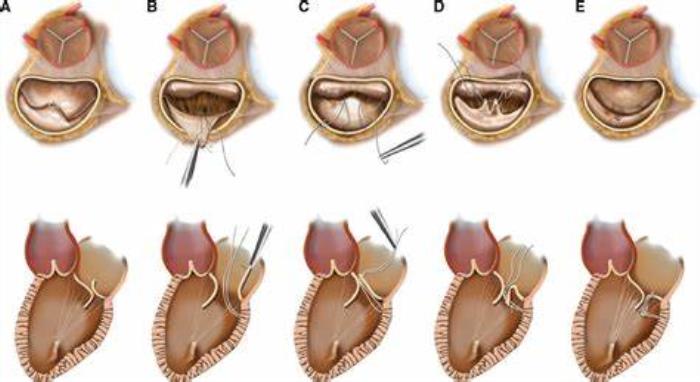
Echocardiography is crucial in planning for mitral valve surgery or repair. By providing detailed images of the mitral valve’s structure, blood flow patterns, and surrounding heart tissues, it helps surgeons determine the most appropriate surgical approach. It also aids in measuring the size of the valve or the heart chambers, ensuring that the correct surgical technique is chosen for the best possible outcome.
Limitations of Echocardiography in Diagnosing Mitral Valve Disease
While echocardiography is a powerful diagnostic tool, it has limitations. Its effectiveness can be reduced in certain patients, such as those who are obese or have lung disease, as these factors can interfere with sound wave transmission. Additionally, echocardiography may not provide as detailed information about very small or subtle valve abnormalities, which may require further diagnostic tests such as cardiac MRI or a transesophageal echocardiogram for more precise evaluation.
The Role of 3D Echocardiography in Advanced Diagnosis of Mitral Valve Disorders
3D echocardiography provides an advanced approach to diagnosing mitral valve disorders by offering three-dimensional images of the heart and its structures. This technique allows for a more accurate assessment of the mitral valve’s shape, size, and function, which is particularly useful in planning complex surgical repairs. By providing a clearer and more detailed picture of the valve, 3D echocardiography can help tailor individualized treatment plans for patients.
Combining Echocardiography with Other Diagnostic Tests for a Complete Picture
To obtain a comprehensive understanding of mitral valve disease, echocardiography is often combined with other diagnostic tests. For example, a cardiac MRI or CT scan may be used to assess heart anatomy and valve structure in more detail. When combined with laboratory tests, such as BNP (brain natriuretic peptide) levels, this multi-modal approach provides a complete picture of the disease, helping clinicians make more informed treatment decisions.
How Echocardiography is Used Post-Surgery to Monitor Mitral Valve Function
Post-surgery, echocardiography is used to monitor the function of the mitral valve and ensure that the surgery has been successful. It helps detect any complications, such as residual valve regurgitation, or problems like clot formation or infection. Regular echocardiograms after surgery ensure that any issues are identified early, allowing for timely interventions if necessary.
Patient Preparation for an Echocardiogram: What to Expect
Preparing for an echocardiogram typically involves minimal steps. Patients may be asked to avoid eating or drinking for a few hours before the test, especially if a transesophageal echocardiogram is planned. During the procedure, the patient will lie on their back while a gel is applied to the chest, and a transducer is moved over the area to capture images. The procedure is painless, and patients can resume normal activities shortly after.
Latest Advancements in Mitral Valve Disease Treatment
Explore the latest advancements in mitral valve disease treatment. This section covers innovative approaches, including minimally invasive techniques and new medical therapies aimed at improving patient outcomes.
Conclusion: The Value of Echocardiography in Early Detection and Management of Mitral Valve Disease
Echocardiography is invaluable in both the early detection and ongoing management of mitral valve disease. It provides detailed insights into the condition of the mitral valve and surrounding heart structures, enabling early diagnosis and treatment. By offering non-invasive, real-time imaging, echocardiography plays a central role in improving patient outcomes, guiding treatment decisions, and monitoring post-surgery recovery.
Best Mitral Valve Replacement Surgery in India
The Best Mitral Valve Replacement Surgery in India is designed to restore heart function in patients with mitral valve disease, providing effective solutions to improve quality of life and heart health.
Best Mitral Valve Replacement Surgeons in India
The Best Mitral Valve Replacement Surgeons in India are skilled in valve replacement techniques, providing personalized care to achieve successful outcomes for patients with mitral valve conditions.
FAQ:
How does an echocardiogram diagnose mitral valve disease?
An echocardiogram uses sound waves to create images of the heart, allowing doctors to assess the mitral valve's structure and function, identifying issues like regurgitation or stenosis.
What is the difference between transthoracic and transesophageal echocardiograms?
Transthoracic echocardiography is performed externally on the chest, while transesophageal echocardiography involves inserting a probe into the esophagus for clearer images of the heart.
Can echocardiography detect mitral valve regurgitation or stenosis?
Yes, echocardiography can detect both mitral valve regurgitation (leakage) and stenosis (narrowing), helping determine the severity of the condition.
How often should I have an echocardiogram if I have mitral valve disease?
The frequency depends on the severity of the disease and your doctor’s recommendation, but it’s often done annually or more frequently if the disease is progressing.
Is echocardiography the most accurate method for diagnosing mitral valve problems?
Echocardiography is highly accurate for diagnosing mitral valve disease, but additional imaging techniques may be used for more detailed analysis in complex cases.