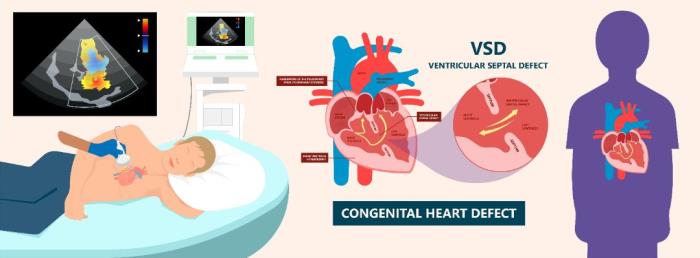
Accurate imaging is crucial in diagnosing and assessing Ventricular Septal Defect (VSD), as it helps determine the size, location, and impact of the defect on heart function. Early diagnosis through advanced imaging techniques enables timely intervention, which is essential for preventing complications such as heart failure and pulmonary hypertension. With ongoing advancements in diagnostic imaging, healthcare providers can offer more precise and effective treatment options for individuals with VSD.
What is Ventricular Septal Defect (VSD) and Why Early Diagnosis Matters
Ventricular Septal Defect (VSD) is a congenital heart condition where there is a hole in the septum separating the left and right ventricles of the heart. If left untreated, VSD can lead to increased blood flow to the lungs, causing pulmonary overcirculation, heart failure, and delayed growth in children. Early diagnosis is crucial to prevent these long-term complications and to guide the best course of treatment, including surgical or catheter-based interventions.
Traditional Imaging Techniques Used to Diagnose VSD
Traditionally, doctors have relied on basic imaging techniques such as chest X-rays and electrocardiograms (ECG) to diagnose VSD. These tools provide initial insights into heart structure and function but are often insufficient for detailed evaluation. A chest X-ray may reveal an enlarged heart, which is a sign of VSD, but it does not show the defect itself. Similarly, an ECG can detect abnormal heart rhythms resulting from VSD, but it cannot pinpoint the exact location or size of the hole.

Advancements in Echocardiography for VSD Detection
Echocardiography has become the gold standard for diagnosing VSD due to its ability to produce real-time images of the heart’s structures. It allows doctors to assess the size and location of the VSD, as well as the impact of the defect on heart function. Doppler ultrasound, a feature of echocardiography, further enhances the ability to visualize blood flow across the defect, providing valuable information about the severity of the condition.
3D Echocardiography: Enhancing VSD Visualization
Advancements in 3D echocardiography have revolutionized the ability to visualize VSDs, especially in complex cases. Unlike traditional 2D imaging, 3D echocardiography provides a more comprehensive view of the heart, allowing clinicians to evaluate the defect from multiple angles. This improved visualization aids in the precise assessment of the VSD’s size, location, and its effect on heart function, which is crucial for planning surgery or other interventions.
The Role of Cardiac MRI in Diagnosing Complex VSDs
For more complex cases of VSD, where echocardiography may not provide enough detail, cardiac MRI is often used. MRI provides high-resolution images of the heart’s structures, including the septum, and offers detailed information about blood flow dynamics. It is particularly useful in assessing VSDs that are difficult to visualize with other imaging modalities, allowing for more accurate diagnosis and surgical planning.
How CT Scans are Used in VSD Diagnosis and Surgical Planning
Cardiac CT scans are less commonly used for initial diagnosis but can be very helpful in surgical planning, especially in older patients or those with complex congenital defects. CT imaging provides detailed, 3D images of the heart and vessels, allowing surgeons to visualize the precise anatomy of the VSD and surrounding structures. This information is invaluable for planning the most effective surgical approach, particularly in cases where traditional echocardiography or MRI may not provide enough clarity.
Doppler Ultrasound: A Vital Tool for Assessing Blood Flow in VSD
Doppler ultrasound plays a crucial role in assessing blood flow across a ventricular septal defect (VSD). It helps identify the direction, velocity, and volume of blood flow through the defect, which is essential for evaluating the severity of the condition and planning treatment. This non-invasive imaging technique provides real-time data that is vital for making informed decisions about surgery or other interventions.
The Benefits of Using High-Resolution Imaging in VSD Diagnosis
High-resolution imaging, such as advanced echocardiography, allows for detailed visualization of the heart’s structures, enabling more accurate detection of VSDs. By providing clear images of the ventricular septum and surrounding heart tissue, high-resolution imaging enhances diagnostic accuracy and helps doctors determine the size and location of the defect, which are crucial for treatment planning.
Real-Time Imaging for Monitoring VSD Progression
Real-time imaging, like Doppler ultrasound or 3D echocardiography, allows healthcare providers to monitor the progression of VSD over time. This dynamic monitoring helps track changes in the size of the defect, blood flow, and the development of complications, offering a more comprehensive understanding of the patient’s condition and guiding timely interventions.
The Role of Contrast Agents in VSD Imaging
Contrast agents, often used in conjunction with echocardiography or cardiac MRI, enhance the visualization of blood flow and heart structures. In VSD diagnosis, contrast agents help to highlight blood flow across the defect more clearly, especially in cases where the VSD is small or difficult to see, improving diagnostic precision and aiding in the assessment of the defect’s severity.

Combining Imaging Techniques for Comprehensive VSD Diagnosis
Using a combination of imaging techniques, such as echocardiography, Doppler ultrasound, and cardiac MRI, provides a comprehensive view of VSD. This multimodal approach allows for detailed assessment of the defect’s size, location, and impact on heart function, leading to more accurate diagnosis and better-informed decisions regarding surgery or other treatments.
Artificial Intelligence in VSD Diagnosis: Emerging Trends
Artificial intelligence (AI) is increasingly being integrated into VSD diagnosis to analyze imaging data more efficiently and accurately. AI algorithms can detect subtle patterns in imaging scans that may be overlooked by the human eye, helping to identify VSDs earlier and more reliably. This technology holds promise for enhancing diagnostic precision and reducing human error in clinical settings.
The Role of Imaging in Pre-Surgical Planning for VSD Repair
Imaging plays a critical role in pre-surgical planning for VSD repair by providing detailed maps of the heart’s structures. It helps surgeons understand the size, location, and characteristics of the defect, which is essential for selecting the most appropriate surgical approach. Advanced imaging techniques also help identify any associated abnormalities that may require simultaneous treatment.
How Advances in Imaging Have Reduced Diagnostic Errors in VSD
Advances in imaging, such as 3D echocardiography and high-resolution MRI, have significantly reduced diagnostic errors in VSD. These technologies provide clearer, more accurate images, allowing for better identification of small or complex defects that might have been missed with older methods. This leads to more precise diagnosis and treatment planning, ultimately improving patient outcomes.
The Use of Imaging in Post-Surgery Monitoring of VSD
Post-surgery monitoring of VSD through imaging is essential to ensure the defect is fully repaired and to detect any complications, such as residual defects or leakage. Imaging techniques like echocardiography and cardiac MRI are regularly used to assess the heart’s function and check for any signs of recurrent issues, allowing for prompt intervention if necessary.
Benefits of Early VSD Detection Through Advanced Imaging
Early detection of VSD through advanced imaging techniques can lead to timely interventions, preventing complications such as heart failure, pulmonary hypertension, and arrhythmias. By identifying the defect before it causes significant damage to the heart or lungs, patients can receive treatment early, improving the prognosis and reducing the risk of long-term complications.
The Future of VSD Imaging: What’s on the Horizon?
The future of VSD imaging is poised to benefit from advancements in technologies like AI, improved 3D imaging, and more precise contrast agents. These innovations will likely lead to even earlier detection, better surgical planning, and enhanced monitoring of post-surgery recovery. The goal is to make VSD diagnosis and management more accurate, efficient, and accessible, further improving patient outcomes.
Cost and Accessibility of Advanced Imaging Techniques for VSD
While advanced imaging techniques, such as cardiac MRI and 3D echocardiography, provide highly detailed images, they can be costly and may not be widely accessible in all healthcare settings. However, as technology advances and becomes more widespread, the cost of these techniques is expected to decrease, making them more accessible to a broader range of patients and healthcare facilities.
Conclusion: Why Advanced Imaging is Essential for Effective VSD Management
Advanced imaging techniques are indispensable for the effective diagnosis, treatment planning, and monitoring of VSD. They provide accurate, detailed insights into the heart’s structure and function, allowing for early detection, precise treatment, and better long-term management. With continued advancements, these imaging tools will play an even greater role in improving patient outcomes and enhancing the overall care of individuals with VSD.
Best VSD Surgery in India
The Best VSD Surgery in India offers effective treatment for ventricular septal defects, helping improve heart function and quality of life for patients with congenital heart defects.
Best VSD Surgery Hospitals in India
The best vsd surgery hospitals in india are equipped with advanced cardiac care facilities and specialized pediatric teams, providing comprehensive care from diagnosis through recovery.
VSD Surgery Cost in India
The vsd surgery cost in india is affordable and transparent, offering high-quality care and accessible options for patients at leading hospitals.
Best VSD Surgeons in India
The Best VSD Surgeons in India are experts in congenital heart defect repairs, providing precise and compassionate care tailored to each patient’s unique needs.
FAQ
How does echocardiography help in diagnosing VSD?
Echocardiography uses sound waves to create detailed images of the heart. It can identify the presence of a VSD, assess its size and location, and evaluate the impact on heart function.
What are the benefits of using 3D echocardiography for VSD detection?
3D echocardiography provides a more comprehensive, detailed view of the heart’s structures, allowing for more accurate assessment of VSD size and complexity compared to traditional 2D imaging.
Can cardiac MRI be used for diagnosing all types of VSD?
Cardiac MRI is effective for diagnosing most types of VSD, particularly complex or large defects, by providing high-resolution images of the heart and great vessels.
How accurate are modern imaging techniques in diagnosing VSD?
Modern imaging techniques, including echocardiography, Doppler ultrasound, and cardiac MRI, are highly accurate in diagnosing VSD, with advanced methods offering greater precision in detecting small or complex defects.
Why is real-time imaging important for monitoring VSD progression?
Real-time imaging, such as Doppler ultrasound, allows for continuous monitoring of blood flow across the VSD, providing immediate insights into any changes in the defect’s size, severity, or impact on heart function, guiding timely treatment.